AutoPET Challenge 2022: Step-by-Step Lesion Segmentation in Whole-body FDG-PET/CT
Publication
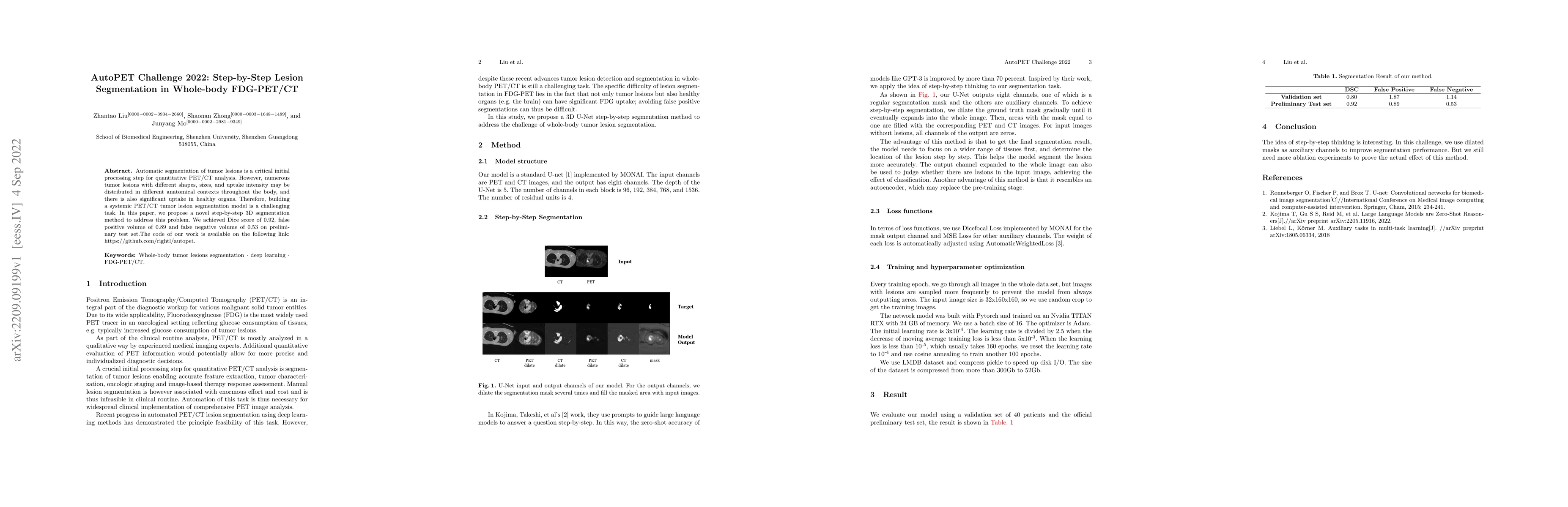
Metrics
AI Quick Summary
The paper proposes a novel step-by-step 3D segmentation method for automatic tumor lesion segmentation in whole-body FDG-PET/CT scans, achieving a Dice score of 0.92, with false positive and negative volumes of 0.89 and 0.53 respectively. The method addresses the complexity of segmenting lesions with varying shapes, sizes, and intensities amidst healthy organ uptake.
Paper Preview
Abstract
Automatic segmentation of tumor lesions is a critical initial processing step for quantitative PET/CT analysis. However, numerous tumor lesions with different shapes, sizes, and uptake intensity may be distributed in different anatomical contexts throughout the body, and there is also significant uptake in healthy organs. Therefore, building a systemic PET/CT tumor lesion segmentation model is a challenging task. In this paper, we propose a novel step-by-step 3D segmentation method to address this problem. We achieved Dice score of 0.92, false positive volume of 0.89 and false negative volume of 0.53 on preliminary test set.The code of our work is available on the following link: https://github.com/rightl/autopet.
AI Key Findings
Get AI-generated insights about this paper's methodology, results, significance, and more — seven facets brought into focus.
Impact
Paper Details
Authors
PDF Preview
Key Terms
Citation Network
Current paper (gray), citations (green), references (blue)
Display is limited for performance on very large graphs.
Discussion 0