Authors
Summary
Automated segmentation of cancerous lesions in PET/CT scans is a crucial first step in quantitative image analysis. However, training deep learning models for segmentation with high accuracy is particularly challenging due to the variations in lesion size, shape, and radiotracer uptake. These lesions can appear in different parts of the body, often near healthy organs that also exhibit considerable uptake, making the task even more complex. As a result, creating an effective segmentation model for routine PET/CT image analysis is challenging. In this study, we utilized a 3D Residual UNet model and employed the Generalized Dice Focal Loss function to train the model on the AutoPET Challenge 2024 dataset. We conducted a 5-fold cross-validation and used an average ensembling technique using the models from the five folds. In the preliminary test phase for Task-1, the average ensemble achieved a mean Dice Similarity Coefficient (DSC) of 0.6687, mean false negative volume (FNV) of 10.9522 ml and mean false positive volume (FPV) 2.9684 ml. More details about the algorithm can be found on our GitHub repository: https://github.com/ahxmeds/autosegnet2024.git. The training code has been shared via the repository: https://github.com/ahxmeds/autopet2024.git.
AI Key Findings
Generated Sep 03, 2025
Methodology
The research utilized a 3D Residual UNet model, trained with Generalized Dice Focal Loss on the AutoPET Challenge 2024 dataset. A 5-fold cross-validation was conducted, and an average ensembling technique was applied using models from the five folds.
Key Results
- The average ensemble achieved a mean Dice Similarity Coefficient (DSC) of 0.6687, mean false negative volume (FNV) of 10.9522 ml, and mean false positive volume (FPV) of 2.9684 ml in the preliminary test phase for Task-1.
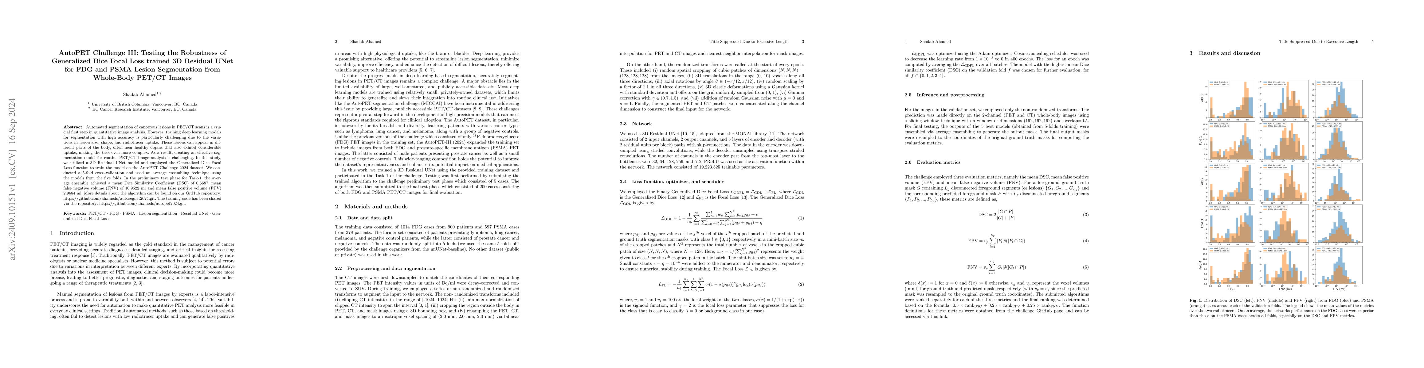
- Network performance on FDG cases was superior to PSMA cases, particularly in DSC and FPV metrics, across all validation folds.
Significance
This study aims to develop an effective segmentation model for routine PET/CT image analysis, which is crucial for quantitative image analysis in cancer diagnosis and treatment planning.
Technical Contribution
The paper presents a deep Residual UNet model trained with Generalized Dice Focal Loss for automated lesion segmentation in FDG and PSMA PET/CT images, demonstrating a 5-fold cross-validation approach.
Novelty
This work introduces the use of Generalized Dice Focal Loss in a 3D Residual UNet model for robust lesion segmentation in PET/CT images, addressing the challenges posed by variations in lesion characteristics and radiotracer uptake.
Limitations
- The study was limited by the variations in lesion size, shape, and radiotracer uptake, which pose challenges in training deep learning models for accurate segmentation.
- Performance discrepancies between FDG and PSMA cases suggest potential room for improvement in handling diverse lesion characteristics.
Future Work
- Enhancing the objective function by incorporating terms for False Positive Volume (FPV) and False Negative Volume (FNV) to better penalize predictions with high FPV or FNV.
- Investigating the impact of additional lesion-level and patient-level lesion measures on segmentation network performance.
Paper Details
PDF Preview
Citation Network
Current paper (gray), citations (green), references (blue)
Display is limited for performance on very large graphs.
Similar Papers
Found 4 papersGeneralized Dice Focal Loss trained 3D Residual UNet for Automated Lesion Segmentation in Whole-Body FDG PET/CT Images
Arman Rahmim, Shadab Ahamed
Dual channel CW nnU-Net for 3D PET-CT Lesion Segmentation in 2024 autoPET III Challenge
Ching-Wei Wang, Ting-Sheng Su, Keng-Wei Liu
Lesion Segmentation in Whole-Body Multi-Tracer PET-CT Images; a Contribution to AutoPET 2024 Challenge
Simone Bendazzoli, Mehdi Astaraki
Autopet III challenge: Incorporating anatomical knowledge into nnUNet for lesion segmentation in PET/CT
Constantin Seibold, Ken Herrmann, Jens Kleesiek et al.
No citations found for this paper.
Comments (0)