ArxivLens

Quick Summary:

This paper proposes a Bayesian-based method for aberration correction and numerical diffraction to enhance lensfree on-chip microscopy, addressing interferometric aberrations, acquisition noise, and reconstruction artifacts. Experimental results show a 25% improvement in numerical aperture and a 2.3 dB to 3.8 dB increase in signal-to-noise ratio compared to standard methods.

Journal Reference: Optics Letters Vol. 40, Issue 10, pp. 2233-2236 (2015)
DOI: 10.1364/OL.40.002233
arXiv Id: 1503.00046
Comments: Optics Letters, vol. 40, no. 10, pp. 2233-2236 (2015) https://www.osapublishing.org/ol/abstract.cfm?uri=ol-40-10-2233
Date Published: 2015-05-11
Date Updated: 2015-05-11

Categories:

Key Terms (TF-IDF):

reconstructed (0.270)
reconstructed (0.268)
snr (0.257)
chip (0.245)
chip (0.235)
db (0.229)
snr (0.227)
microscopy (0.222)
na (0.221)
improvements (0.199)
microscopy (0.199)
image (0.190)
issues (0.186)
improvements (0.180)
image (0.177)
imaging (0.174)
issues (0.167)
imaging (0.165)
method (0.161)
noise (0.156)
License: http://arxiv.org/licenses/nonexclusive-distrib/1.0/

Summary:

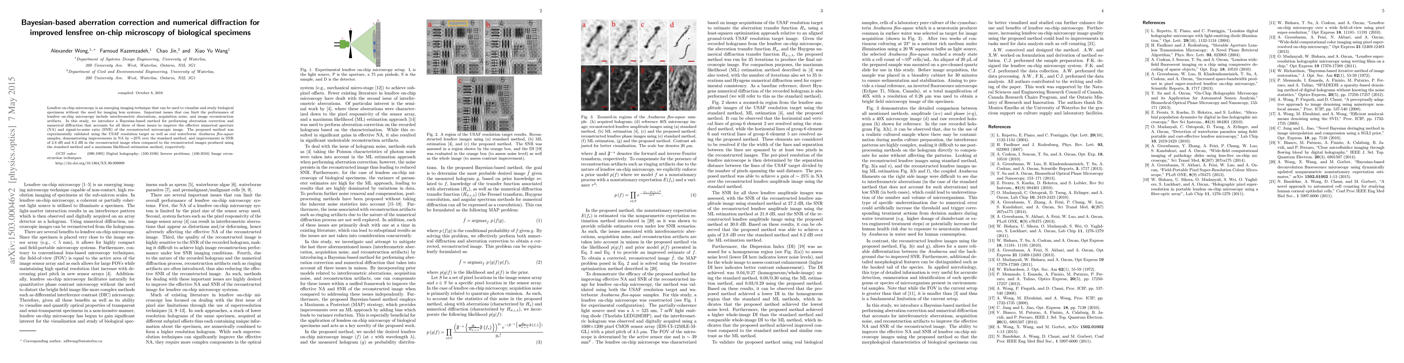
Lensfree on-chip microscopy is an emerging imaging technique that can be used to visualize and study biological specimens without the need for imaging lens systems. Important issues that can limit the performance of lensfree on-chip microscopy include interferometric aberrations, acquisition noise, and image reconstruction artifacts. In this study, we introduce a Bayesian-based method for performing aberration correction and numerical diffraction that accounts for all three of these issues to improve the effective numerical aperture (NA) and signal-to-noise ratio (SNR) of the reconstructed microscopic image. The proposed method was experimentally validated using the USAF resolution target as well as real waterborne Anabaena flos-aquae samples, demonstrating improvements in NA by ~25% over the standard method, and improvements in SNR of 2.3 dB and 3.8 dB in the reconstructed image when compared to the reconstructed images produced using the standard method and a maximum likelihood estimation method, respectively.

Citation & Reference Network Visualization

This graph shows how this paper is connected to other research. The current paper is shown in dark gray, papers that cite it are in green, and papers it references are in blue. You can:

Current Paper
Papers citing this work (Citations)
Papers cited by this work (References)

• Hover over any node to see the paper title

• Click a node to view that paper's details

• Drag nodes to rearrange the graph

• Use the zoom controls (bottom right) to zoom in/out

Citations and References

Title Authors Year Actions
Advancing the science of dynamic airborne nanosized particles using Nano-DIHM
D. Pal, Y. Nazarenko, T. Preston, P. Ariya
2021
View Paper
Enhanced spectral lightfield fusion microscopy via deep computational optics for whole-slide pathology
Farnoud Kazemzadeh, H. Li-Chang, A. Wong
2019
View Paper
Lensless digital holographic microscopy and its applications in biomedicine and environmental monitoring.
Yichen Wu, A. Ozcan
2017
View Paper
Laser Light-field Fusion for Wide-field Lensfree On-chip Phase Contrast Microscopy of Nanoparticles
Farnoud Kazemzadeh, A. Wong
2016
View Paper
Compact, Field-Portable Lens-free Microscope using Superresolution Spatio-Spectral Light-field Fusion
Farnoud Kazemzadeh, E. Kuang, A. Wong
2016
View Paper
Laser light-field fusion for wide-field lensfree on-chip phase contrast nanoscopy
Farnoud Kazemzadeh, A. Wong
2016
View Paper
Simultaneous Multispectral Imaging: Using Multiview Computational Compressive Sensing
Farnoud Kazemzadeh
2016
View Paper
Lens-free Multi-Laser Spectral Light-Field Fusion Microscopy
Farnoud Kazemzadeh, A. Wong
2015
View Paper
Multispectral digital holographic microscopy with applications in water quality assessment
Farnoud Kazemzadeh, Chao Jin, Mei Yu, Robert Amelard, Shahid A. Haider, S. Saini, M. Emelko, David A Clausi, A. Wong
2015
View Paper
Lens-free spectral light-field fusion microscopy for contrast- and resolution-enhanced imaging of biological specimens.
Farnoud Kazemzadeh, Chao Jin, Sara Molladavoodi, Yushan Mei, M. Emelko, M. Gorbet, A. Wong
2015
View Paper
A Bayesian Residual Transform for Signal Processing
A. Wong, Xiaoyu Wang
2014
View Paper
Digital Holographic Microscopy
Farnoud Kazemzadeh, A. Wong
2019
View Paper

Similar Papers

Fourier-Based 3D Multistage Transformer for Aberration Correction in Multicellular Specimens

Srigokul Upadhyayula, Evan Huang, Thayer Alshaabi, Gaoxiang Liu, Daniel E. Milkie, Cyna Shirazinejad, Jason L. Hong, Kemal Achour, Frederik Görlitz, Ana Milunovic-Jevtic, Cat Simmons, Ibrahim S. Abuzahriyeh, Erin Hong, Samara Erin Williams, Nathanael Harrison, Eun Seok Bae, Alison N. Killilea, David G. Drubin, Ian A. Swinburne, Eric Betzig

Published: 2025-03-16

BEACON -- Automated Aberration Correction for Scanning Transmission Electron Microscopy using Bayesian Optimization

Stephanie M. Ribet, Georgios Varnavides, Colin Ophus, Peter Ercius, Kunwoo Park, Alexander J. Pattison, Jungwon Park, Marcus M. Noack, Earl Kirkland

Published: 2024-10-18

Comments (0)

This website uses cookies to ensure you get the best experience. Learn more