BEFD: Boundary Enhancement and Feature Denoising for Vessel Segmentation
Publication
Metrics
AI Quick Summary
A new module called Boundary Enhancement and Feature Denoising (BEFD) improves blood vessel segmentation by enhancing boundary information and reducing noise in medical images, leading to superior performance on retinal vessel and angiocarpy datasets.
Paper Preview
Abstract
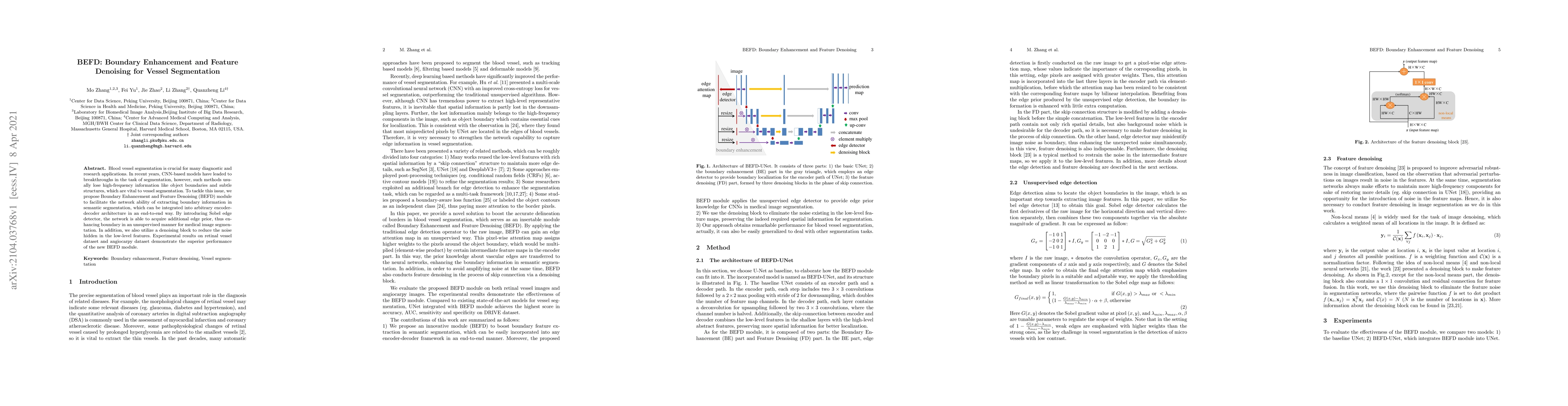
Blood vessel segmentation is crucial for many diagnostic and research applications. In recent years, CNN-based models have leaded to breakthroughs in the task of segmentation, however, such methods usually lose high-frequency information like object boundaries and subtle structures, which are vital to vessel segmentation. To tackle this issue, we propose Boundary Enhancement and Feature Denoising (BEFD) module to facilitate the network ability of extracting boundary information in semantic segmentation, which can be integrated into arbitrary encoder-decoder architecture in an end-to-end way. By introducing Sobel edge detector, the network is able to acquire additional edge prior, thus enhancing boundary in an unsupervised manner for medical image segmentation. In addition, we also utilize a denoising block to reduce the noise hidden in the low-level features. Experimental results on retinal vessel dataset and angiocarpy dataset demonstrate the superior performance of the new BEFD module.
AI Key Findings
Get AI-generated insights about this paper's methodology, results, significance, and more — seven facets brought into focus.
Impact
Paper Details
Authors
PDF Preview
Key Terms
Citation Network
Current paper (gray), citations (green), references (blue)
Display is limited for performance on very large graphs.
Discussion 0