Beyond the Eye: A Relational Model for Early Dementia Detection Using Retinal OCTA Images
Publication
Metrics
AI Quick Summary
This paper introduces PolarNet+, a novel model using retinal OCTA images to detect early-onset Alzheimer's disease (EOAD) and mild cognitive impairment (MCI) by mapping OCTA images into polar coordinates for ETDRS grid analysis, employing a multi-view module for comprehensive image analysis, and utilizing a regional relationship module to identify eye-brain connections and discriminative patterns for dementia detection.
Paper Preview
Abstract
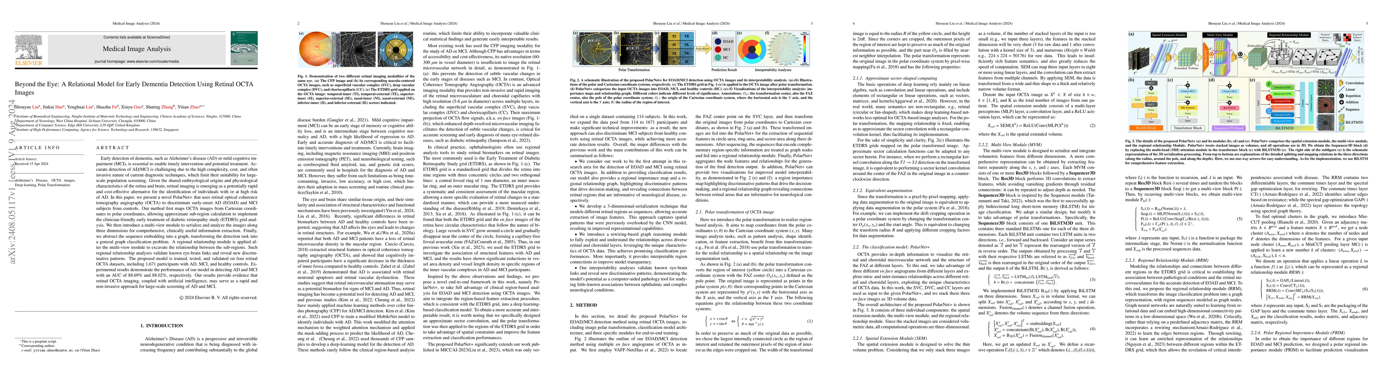
Early detection of dementia, such as Alzheimer's disease (AD) or mild cognitive impairment (MCI), is essential to enable timely intervention and potential treatment. Accurate detection of AD/MCI is challenging due to the high complexity, cost, and often invasive nature of current diagnostic techniques, which limit their suitability for large-scale population screening. Given the shared embryological origins and physiological characteristics of the retina and brain, retinal imaging is emerging as a potentially rapid and cost-effective alternative for the identification of individuals with or at high risk of AD. In this paper, we present a novel PolarNet+ that uses retinal optical coherence tomography angiography (OCTA) to discriminate early-onset AD (EOAD) and MCI subjects from controls. Our method first maps OCTA images from Cartesian coordinates to polar coordinates, allowing approximate sub-region calculation to implement the clinician-friendly early treatment of diabetic retinopathy study (ETDRS) grid analysis. We then introduce a multi-view module to serialize and analyze the images along three dimensions for comprehensive, clinically useful information extraction. Finally, we abstract the sequence embedding into a graph, transforming the detection task into a general graph classification problem. A regional relationship module is applied after the multi-view module to excavate the relationship between the sub-regions. Such regional relationship analyses validate known eye-brain links and reveal new discriminative patterns.
AI Key Findings
Get AI-generated insights about this paper's methodology, results, significance, and more — seven facets brought into focus.
Impact
Authors
PDF Preview
Citation Network
Current paper (gray), citations (green), references (blue)
Display is limited for performance on very large graphs.
Discussion 0