ArxivLens

Quick Summary:

This paper introduces a novel method for estimating the contrast transfer function (CTF) in electron microscopy, using the multi-taper method for power spectral density estimation to reduce bias and variance. The method leverages the known properties of the CTF and background to enhance accuracy, achieving precise zero-crossings in the low-mid frequency range.

arXiv Id: 1908.03454
Date Published: 2020-01-29
Date Updated: 2020-01-29

Categories:

Key Terms (TF-IDF):

bands (0.230)
estimation (0.229)
frequency (0.215)
ice (0.169)
novel method (0.162)
frequency range (0.162)
termed (0.160)
power (0.158)
distortion (0.158)
pass (0.156)
lens (0.155)
present novel (0.151)
method (0.146)
causes (0.146)
power spectrum (0.144)
filter (0.143)
mid (0.142)
method based (0.140)
spread (0.139)
projection (0.139)
License: http://arxiv.org/licenses/nonexclusive-distrib/1.0/

Summary:

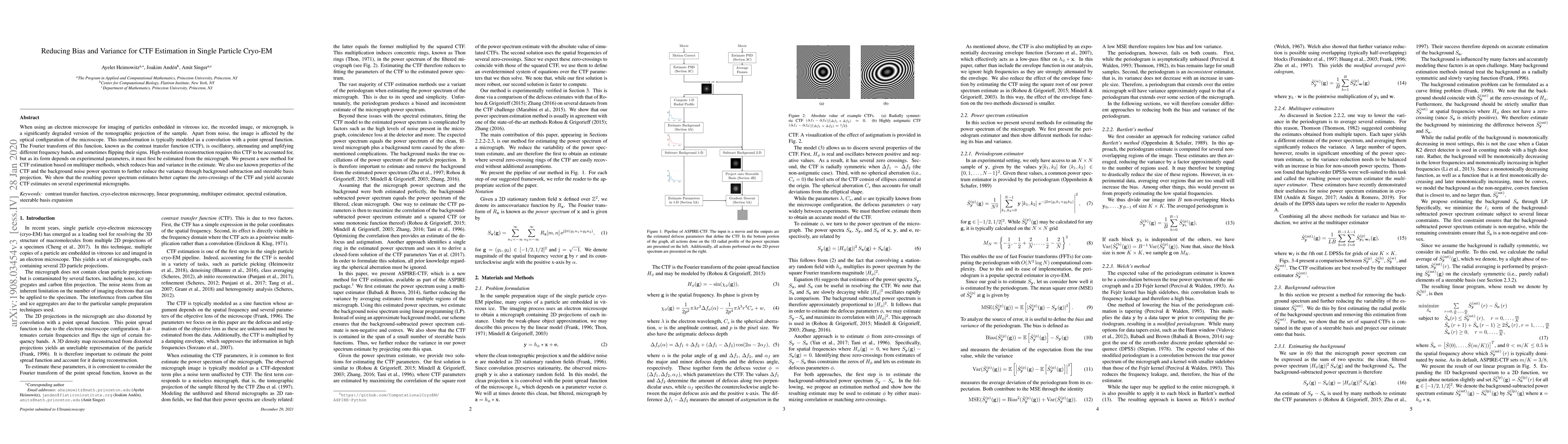
When using an electron microscope for imaging of particles embedded in vitreous ice, the objective lens will inevitably corrupt the projection images. This corruption manifests as a band-pass filter on the micrograph. In addition, it causes the phase of several frequency bands to be flipped and distorts frequency bands. As a precursor to compensating for this distortion, the corrupting point spread function, which is termed the contrast transfer function (CTF) in reciprocal space, must be estimated. In this paper, we will present a novel method for CTF estimation. Our method is based on the multi-taper method for power spectral density estimation, which aims to reduce the bias and variance of the estimator. Furthermore, we use known properties of the CTF and of the background of the power spectrum to increase the accuracy of our estimation. We will show that the resulting estimates capture the zero-crossings of the CTF in the low-mid frequency range.

Citation & Reference Network Visualization

This graph shows how this paper is connected to other research. The current paper is shown in dark gray, papers that cite it are in green, and papers it references are in blue. You can:

Current Paper
Papers citing this work (Citations)
Papers cited by this work (References)

• Hover over any node to see the paper title

• Click a node to view that paper's details

• Drag nodes to rearrange the graph

• Use the zoom controls (bottom right) to zoom in/out

Citations and References

Title Authors Year Actions
Novel Artificial Intelligence-Based Approaches for Ab Initio Structure Determination and Atomic Model Building for Cryo-Electron Microscopy
Megan C DiIorio, A. W. Kulczyk
2023
View Paper
Representing Steerable Bases for cryo-EM in ASPIRE
Christopher Langfield, Joshua Carmichael, Garrett Wright, J. Andén, A. Singer
2022
View Paper
Emerging Themes in CryoEM—Single Particle Analysis Image Processing
J. Vilas, J. Carazo, C. Sorzano
2022
View Paper
Ab-initio Contrast Estimation and Denoising of Cryo-EM Images
Yunpeng Shi, A. Singer
2022
View Paper
Two-Dimensional Multi-Target Detection: An Autocorrelation Analysis Approach
Shay Kreymer, Tamir Bendory
2021
View Paper
Artificial intelligence advances for de novo molecular structure modeling in cryo‐electron microscopy
Dong Si, Andrew Nakamura, Runbang Tang, Haowen Guan, Jie Hou, Ammaar Firozi, Renzhi Cao, Kyle Hippe, Minglei Zhao
2021
View Paper
Toward single particle reconstruction without particle picking: Breaking the detection limit
Tamir Bendory, Nicolas Boumal, W. Leeb, Eitan Levin, A. Singer
2018
View Paper

Comments (0)

This website uses cookies to ensure you get the best experience. Learn more