Bio-Mechanical Model of the Brain for a Per-Operative Image-Guided Neuronavigator Compensating for "Brain-Shift" Deformations
Publication
Metrics
AI Quick Summary
This paper proposes a bio-mechanical model for a neuronavigator that compensates for "brain-shift" during neurosurgery, generating patient-specific finite element meshes to account for tissue deformations and surgical modifications, thereby improving the accuracy of intra-operative image guidance.
Paper Preview
Abstract
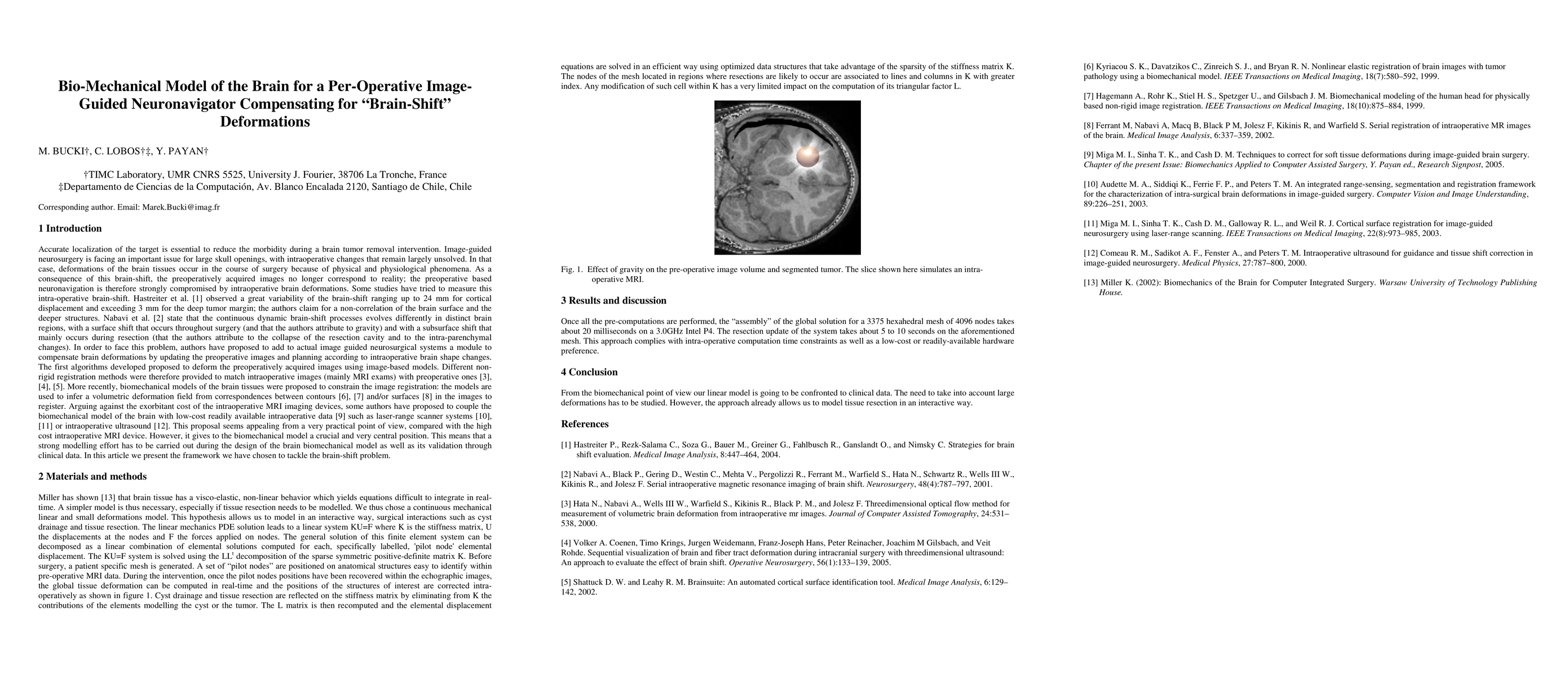
In this paper we present a methodology to address the problem of brain tissue deformation referred to as 'brain-shift'. This deformation occurs throughout a neurosurgery intervention and strongly alters the accuracy of the neuronavigation systems used to date in clinical routine which rely solely on pre-operative patient imaging to locate the surgical target, such as a tumour or a functional area. After a general description of the framework of our intra-operative image-guided system, we describe a procedure to generate patient specific finite element meshes of the brain and propose a biomechanical model which can take into account tissue deformations and surgical procedures that modify the brain structure, like tumour or tissue resection.
AI Key Findings
Get AI-generated insights about this paper's methodology, results, significance, and more — seven facets brought into focus.
Impact
Paper Details
PDF Preview
Key Terms
Citation Network
Current paper (gray), citations (green), references (blue)
Display is limited for performance on very large graphs.
Discussion 0