Biological Tissue Imaging with a Position and Time Sensitive Pixelated Detector
Publication
Metrics
AI Quick Summary
This paper presents a highly parallel, active pixel detector for large-area mass spectrometric imaging of biological tissue sections, achieving high spatial resolution and resolving power. The Timepix detector combined with ion microscope technology enables automated, large-area imaging with significant potential for fast, detailed bio-molecular mass spectrometry.
Paper Preview
Abstract
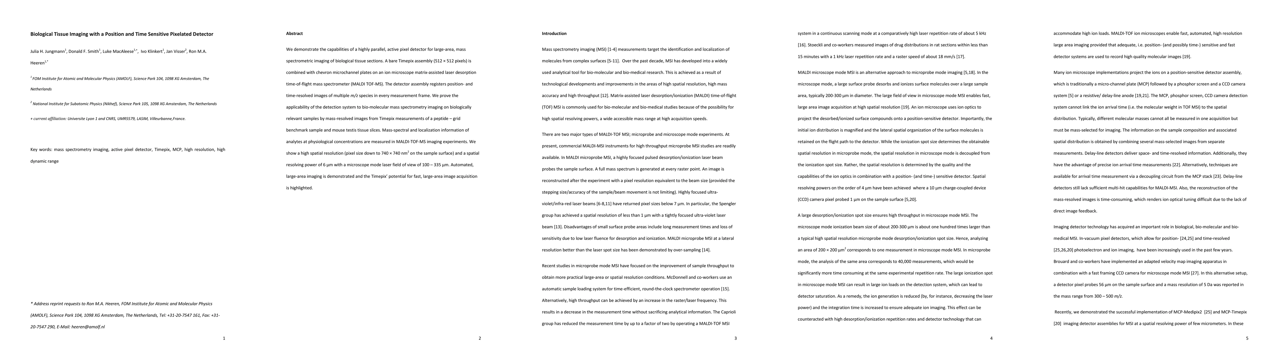
We demonstrate the capabilities of a highly parallel, active pixel detector for large-area, mass spectrometric imaging of biological tissue sections. A bare Timepix assembly (512x512 pixels) is combined with chevron microchannel plates on an ion microscope matrix-assisted laser desorption time-of-flight mass spectrometer (MALDI TOF-MS). The detector assembly registers position- and time-resolved images of multiple m/z species in every measurement frame. We prove the applicability of the detection system to bio-molecular mass spectrometry imaging on biologically relevant samples by mass-resolved images from Timepix measurements of a peptide-grid benchmark sample and mouse testis tissue slices. Mass-spectral and localization information of analytes at physiological concentrations are measured in MALDI-TOF-MS imaging experiments. We show a high spatial resolution (pixel size down to 740x740 nm2 on the sample surface) and a spatial resolving power of 6 {\mu}m with a microscope mode laser field of view of 100-335 {\mu}m. Automated, large-area imaging is demonstrated and the Timepix' potential for fast, large-area image acquisition is highlighted.
AI Key Findings
Get AI-generated insights about this paper's methodology, results, significance, and more — seven facets brought into focus.
Impact
Paper Details
PDF Preview
Key Terms
Citation Network
Current paper (gray), citations (green), references (blue)
Display is limited for performance on very large graphs.
Discussion 0