Publication
Metrics
AI Quick Summary
Researchers found that using X-ray free-electron lasers can image biomolecular structures with minimal damage, contradicting previous estimates. Their approach uses a more detailed imaging model derived from optical coherence theory to tolerate electronic damage.
Paper Preview
Abstract
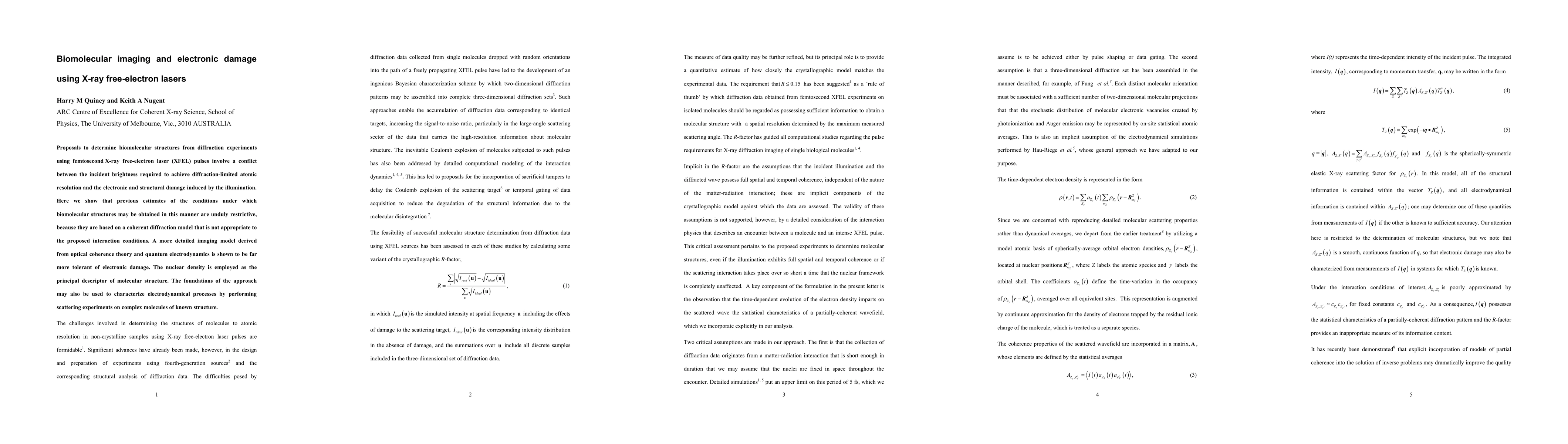
Proposals to determine biomolecular structures from diffraction experiments using femtosecond X-ray free-electron laser (XFEL) pulses involve a conflict between the incident brightness required to achieve diffraction-limited atomic resolution and the electronic and structural damage induced by the illumination. Here we show that previous estimates of the conditions under which biomolecular structures may be obtained in this manner are unduly restrictive, because they are based on a coherent diffraction model that is not appropriate to the proposed interaction conditions. A more detailed imaging model derived from optical coherence theory and quantum electrodynamics is shown to be far more tolerant of electronic damage. The nuclear density is employed as the principal descriptor of molecular structure. The foundations of the approach may also be used to characterize electrodynamical processes by performing scattering experiments on complex molecules of known structure.
AI Key Findings
Get AI-generated insights about this paper's methodology, results, significance, and more — seven facets brought into focus.
Impact
Paper Details
PDF Preview
Key Terms
Citation Network
Current paper (gray), citations (green), references (blue)
Display is limited for performance on very large graphs.
Discussion 0