ArxivLens

Quick Summary:

This paper introduces an unsupervised deep neural network for blind deconvolution in microscopy using cycle consistency and explicit PSF modeling layers, offering a faster and more robust alternative to conventional computationally expensive methods. Experimental results demonstrate the algorithm's effectiveness in improving resolution in widefield fluorescent microscopy.

arXiv Id: 1904.02910
Date Published: 2019-04-08
Date Updated: 2019-04-08

Categories:

Key Terms (TF-IDF):

blind (0.284)
cnn (0.276)
microscopy (0.269)
cnn (0.246)
microscopy (0.243)
layers (0.232)
approaches (0.222)
layers (0.209)
modeling (0.205)
approaches (0.203)
improve (0.190)
modeling (0.188)
improve (0.175)
high performance (0.172)
based approaches (0.168)
extensively (0.162)
high performance (0.161)
computationally expensive (0.160)
algorithm (0.155)
expensive (0.154)
License: http://arxiv.org/licenses/nonexclusive-distrib/1.0/

Summary:

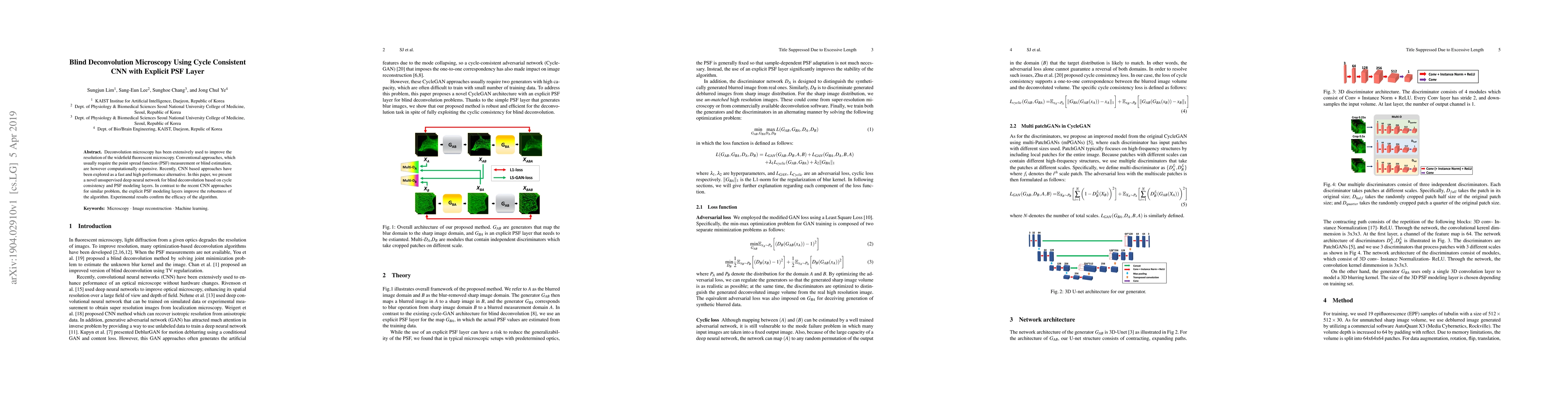
Deconvolution microscopy has been extensively used to improve the resolution of the widefield fluorescent microscopy. Conventional approaches, which usually require the point spread function (PSF) measurement or blind estimation, are however computationally expensive. Recently, CNN based approaches have been explored as a fast and high performance alternative. In this paper, we present a novel unsupervised deep neural network for blind deconvolution based on cycle consistency and PSF modeling layers. In contrast to the recent CNN approaches for similar problem, the explicit PSF modeling layers improve the robustness of the algorithm. Experimental results confirm the efficacy of the algorithm.

Citation & Reference Network Visualization

This graph shows how this paper is connected to other research. The current paper is shown in dark gray, papers that cite it are in green, and papers it references are in blue. You can:

Current Paper
Papers citing this work (Citations)
Papers cited by this work (References)

• Hover over any node to see the paper title

• Click a node to view that paper's details

• Drag nodes to rearrange the graph

• Use the zoom controls (bottom right) to zoom in/out

Citations and References

Title Authors Year Actions
Solving the inverse problem of microscopy deconvolution with a residual Beylkin-Coifman-Rokhlin neural network
Rui Li, M. Kudryashev, Artur Yakimovich
2024
View Paper
Ultrasound image improvement based on parameter optimization of spatially variable point spread function
Jie Zheng, Yifei Chen, Xiang Li, Xin Zhang, Yi Shen
2024
View Paper
Correction of out-of-focus microscopic images by deep learning
Chi Zhang, Hao Jiang, Weihuang Liu, Junyi Li, Shimin Tang, M. Juhas, Yang Zhang
2022
View Paper
High-throughput widefield fluorescence imaging of 3D samples using deep learning for 2D projection image restoration
Edvin Forsgren, Christoffer Edlund, Miniver Oliver, Kalpana Barnes, Rickard Sjögren, Timothy R. Jackson
2022
View Paper
Semi- and Self-Supervised Multi-View Fusion of 3D Microscopy Images using Generative Adversarial Networks
Canyu Yang, Dennis Eschweiler, J. Stegmaier
2021
View Paper
Imaging through scattering media based on semi-supervised learning.
Kaoru Yamazaki, Ryochi Horisaki, J. Tanida
2020
View Paper
Learning to Model and Calibrate Optics Via a Differentiable Wave Optics Simulator
J. Page, P. Favaro
2020
View Paper
CycleGAN With a Blur Kernel for Deconvolution Microscopy: Optimal Transport Geometry
Sungjun Lim, Hyoungjun Park, Sang-Eun Lee, Sunghoe Chang, Byeongsu Sim, J. C. Ye
2019
View Paper
CaGAN: A Cycle-Consistent Generative Adversarial Network With Attention for Low-Dose CT Imaging
Zhiyuan Huang, Zixiang Chen, Qiyang Zhang, Guotao Quan, Min Ji, Cheng-jin Zhang, Yongfeng Yang, Xin Liu, D. Liang, Hairong Zheng, Zhanli Hu
2020
View Paper

Comments (0)

This website uses cookies to ensure you get the best experience. Learn more