This study investigates the relationship between bone density and texture in knee radiographs with minimal versus clinical post-processing in subjects with knee osteoarthritis. It found significant correlations between minimal and clinical post-processing for bone density and varied correlations for texture, with notable differences in bone characteristics between controls and osteoarthritic subjects.
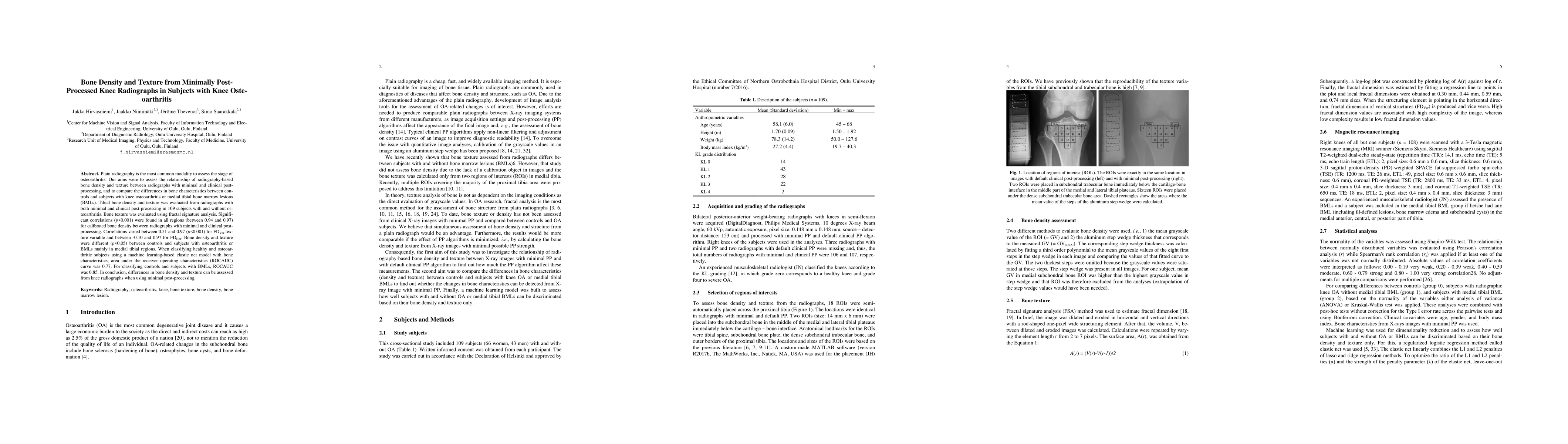
Plain radiography is the most common modality to assess the stage of osteoarthritis. Our aims were to assess the relationship of radiography-based bone density and texture between radiographs with minimal and clinical post-processing, and to compare the differences in bone characteristics between controls and subjects with knee osteoarthritis or medial tibial bone marrow lesions (BMLs). Tibial bone density and texture was evaluated from radiographs with both minimal and clinical post-processing in 109 subjects with and without osteoarthritis. Bone texture was evaluated using fractal signature analysis. Significant correlations (p<0.001) were found in all regions (between 0.94 and 0.97) for calibrated bone density between radiographs with minimal and clinical post-processing. Correlations varied between 0.51 and 0.97 (p<0.001) for FD_Ver texture variable and between -0.10 and 0.97 for FD_Hor. Bone density and texture were different (p<0.05) between controls and subjects with osteoarthritis or BMLs mainly in medial tibial regions. When classifying healthy and osteoarthritic subjects using a machine learning-based elastic net model with bone characteristics, area under the receiver operating characteristics (ROCAUC) curve was 0.77. For classifying controls and subjects with BMLs, ROCAUC was 0.85. In conclusion, differences in bone density and texture can be assessed from knee radiographs when using minimal post-processing.
Get AI-generated insights about this paper's methodology, results, and significance.
Current paper (gray), citations (green), references (blue)
Title | Authors | Year | Actions |
---|---|---|---|
OA-MEN: a fusion deep learning approach for enhanced accuracy in knee osteoarthritis detection and classification using X-Ray imaging
|
Xiaolu Ren, Lingxuan Hou, Shan Liu, Peng Wu, Siming Liang, Haitian Fu, Chengquan Li, Ting Li, Yongjing Cheng
|
2025
|
View Paper |
Computerized diagnosis of knee osteoarthritis from x‐ray images using combined texture features: Data from the osteoarthritis initiative
|
Khadidja Messaoudene, Khaled Harrar
|
2024
|
View Paper |
Artificial intelligence in osteoarthritis detection: a systematic review and meta-analysis.
|
S. Mohammadi, M. Salehi, Ali Jahanshahi, Mohammad Shahrabi Farahani, S. S. Zakavi, Sadra Behrouzieh, Mahdi Gouravani, A. Guermazi
|
2023
|
View Paper |
The developing juvenile distal tibia: Radiographic identification of distinct ontogenetic phases and structural trajectories
|
R. Reid, Catriona Davies, C. Cunningham
|
2022
|
View Paper |
Rapid X-Ray-Based 3-D Finite Element Modeling of Medial Knee Joint Cartilage Biomechanics During Walking
|
S. Jahangir, Ali Mohammadi, M. Mononen, J. Hirvasniemi, J. Suomalainen, S. Saarakkala, R. Korhonen, P. Tanska
|
2022
|
View Paper |
Radiographic interpretation using high-resolution Cbct to diagnose degenerative temporomandibular joint disease
|
J. Bianchi, João Roberto Gonçalves, Antônio Carlos de Oliveira Ruellas, Júlia Vieira Pastana Bianchi, L. Ashman, M. Yatabe, E. Benavides, F. Soki, L. Cevidanes
|
2021
|
View Paper |
Trabecular bone texture analysis of conventional radiographs in the assessment of knee osteoarthritis: review and viewpoint
|
A. Almhdie-Imjabbar, P. Podsiadło, R. Ljuhar, R. Jennane, Khac-Lan Nguyen, H. Toumi, S. Saarakkala, E. Lespessailles
|
2021
|
View Paper |
A machine learning approach to distinguish between knees without and with osteoarthritis using MRI-based radiomic features from tibial bone
|
J. Hirvasniemi, S. Klein, S. Bierma-Zeinstra, M. Vernooij, D. Schiphof, E. Oei
|
2021
|
View Paper |
Discrimination of Low-Energy Acetabular Fractures from Controls Using Computed Tomography-Based Bone Characteristics
|
R. Gebre, J. Hirvasniemi, I. Lantto, S. Saarakkala, J. Leppilahti, T. Jämsä
|
2020
|
View Paper |
Radiographic Image Radiomics Feature Reproducibility: A Preliminary Study on the Impact of Field Size.
|
S. Rastegar, Jalal Beigi, E. Saeedi, Isaac Shiri, Y. Qasempour, M. Rezaei, H. Abdollahi
|
2020
|
View Paper |
Early Detection of Osteoarthritis based on Cartilage Thickness in Knee X-ray Images
|
S. Gornale, P. Patravali
|
2019
|
View Paper |
Adaptive Segmentation of Knee Radiographs for Selecting the Optimal ROI in Texture Analysis
|
N. Bayramoglu, A. Tiulpin, J. Hirvasniemi, M. Nieminen, S. Saarakkala
|
2019
|
View Paper |
Expediting Finite Element Analyses for Subject-Specific Studies of Knee Osteoarthritis: A Literature Review
|
A. Paz, G. Orozco, R. Korhonen
|
2021
|
View Paper |
Deep learning for knee osteoarthritis diagnosis and progression prediction from plain radiographs and clinical data
|
A. Tiulpin
|
2020
|
View Paper |
Radiographic Bone Texture Analysis Using Deep Learning Models for Early Rheumatoid Arthritis Diagnosis
|
Yun-Ju Huang, Miao Shun, K. Zheng, Le Lu, Yuhang Lu, Chihung Lin, C. Kuo
|
2020
|
View Paper |
Comments (0)