ArxivLens

Quick Summary:

This paper proposes a U-net architecture with innovative preprocessing techniques to segment bone regions in low-dose contrast enhanced whole-body CT scans, achieving high mean Dice coefficients across multiple datasets, highlighting the importance of preprocessing for accurate differentiation.

arXiv Id: 2008.05223
Comments: 15 pages, 10 figures and 3 tables. Submitted to The Journal of Physics in Medicine and Biology for possible publication
Date Published: 2020-08-14
Date Updated: 2020-08-14

Key Terms (TF-IDF):

enhanced (0.276)
dose (0.269)
ct (0.265)
enhanced (0.260)
contrast (0.251)
contrast (0.235)
segmentation (0.224)
body (0.218)
regions (0.212)
body (0.209)
regions (0.206)
segmentation (0.203)
difficult (0.195)
difficult (0.181)
achieved (0.176)
achieved (0.169)
intensities (0.155)
segment (0.155)
activation (0.150)
pixel (0.145)
License: http://arxiv.org/licenses/nonexclusive-distrib/1.0/

Summary:

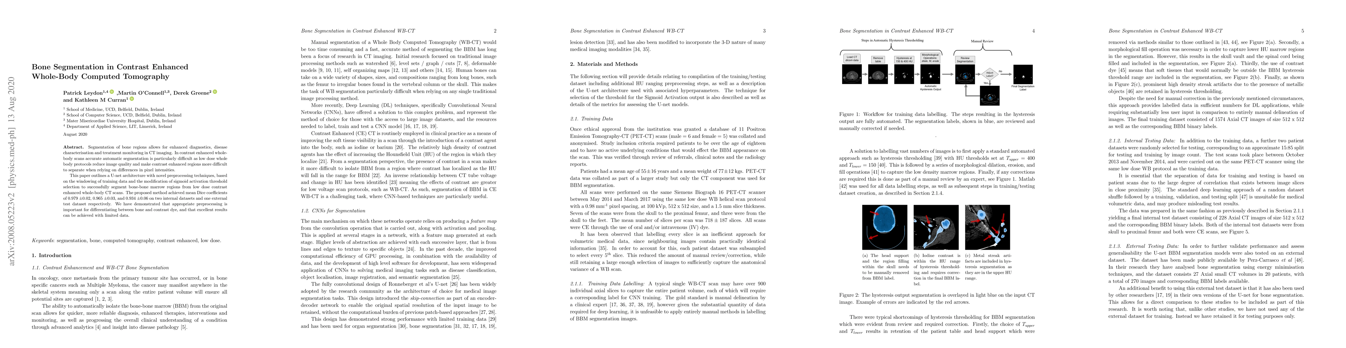
Segmentation of bone regions allows for enhanced diagnostics, disease characterisation and treatment monitoring in CT imaging. In contrast enhanced whole-body scans accurate automatic segmentation is particularly difficult as low dose whole body protocols reduce image quality and make contrast enhanced regions more difficult to separate when relying on differences in pixel intensities. This paper outlines a U-net architecture with novel preprocessing techniques, based on the windowing of training data and the modification of sigmoid activation threshold selection to successfully segment bone-bone marrow regions from low dose contrast enhanced whole-body CT scans. The proposed method achieved mean Dice coefficients of 0.979, 0.965, and 0.934 on two internal datasets and one external test dataset respectively. We have demonstrated that appropriate preprocessing is important for differentiating between bone and contrast dye, and that excellent results can be achieved with limited data.

Citation & Reference Network Visualization

This graph shows how this paper is connected to other research. The current paper is shown in dark gray, papers that cite it are in green, and papers it references are in blue. You can:

Current Paper
Papers citing this work (Citations)
Papers cited by this work (References)

• Hover over any node to see the paper title

• Click a node to view that paper's details

• Drag nodes to rearrange the graph

• Use the zoom controls (bottom right) to zoom in/out

Citations and References

Title Authors Year Actions
Fully Automated Detection and Segmentation Pipeline for the Bone Marrow of the Lytic Bone of Multiple Myeloma Patients
Emmanouil Koutoulakis, Eleftherios Trivizakis, V. Koutoulidis, L. Moulopoulos, E. Terpos, I. Ntanasis-Stathopoulos, P. Malandrakis, Panagiotis Grigoropoulos, Panagiotis Papadopoulos, K. Nikiforaki, Nickolas Papanikolaou, D. Fotiadis, K. Marias
2023
View Paper
Segmentation of bone structures with the use of deep learning techniques
Z. Krawczyk, J. S. ´NSKI, J. Starzyński, Bull. Pol, Ac. Tech
2023
View Paper
Improved distinct bone segmentation in upper-body CT through multi-resolution networks
Eva Schnider, Julia Wolleb, Antal Huck, M. Toranelli, G. Rauter, M. Müller-Gerbl, P. Cattin
2023
View Paper
Ensemble uncertainty as a criterion for dataset expansion in distinct bone segmentation from upper-body CT images
Eva Schnider, Antal Huck, M. Toranelli, G. Rauter, A. Zam, M. Müller-Gerbl, P. Cattin
2022
View Paper
Efficient lower-limb segmentation for large-scale volumetric CT by using projection view and voxel group attention
Fang Chen, Yanting Xie, Peng Xu, Zhe Zhao, Daoqiang Zhang, Hongen Liao
2022
View Paper

Similar Papers

Comments (0)

This website uses cookies to ensure you get the best experience. Learn more