Publication

Published: Jan 13, 2025
Categories: cs.CV

Metrics

Source: ArXiv

Quick Actions

View PDF

AI Quick Summary

This paper proposes a semi-supervised learning framework for sclera segmentation using fewer labeled samples, addressing the scarcity of high-quality datasets. The method integrates domain-specific improvements and spatial transformations to achieve superior segmentation performance.

Quick Answers

What methodology did the authors use?

The research employed a multi-step approach combining machine learning algorithms with traditional image processing techniques to analyze medical images. More in Methodology →

What are the key results?

Improved accuracy in detecting eye diseases from retinal scans — Enhanced object detection capabilities for identifying specific features More in Key Results →

Why is this work significant?

This research has significant implications for the early diagnosis and treatment of eye diseases, improving patient outcomes and quality of life. More in Significance →

What are the main limitations?

Limited dataset size may affect generalizability to diverse populations — Dependence on computational resources for processing large image datasets More in Limitations →

Paper Preview

Abstract

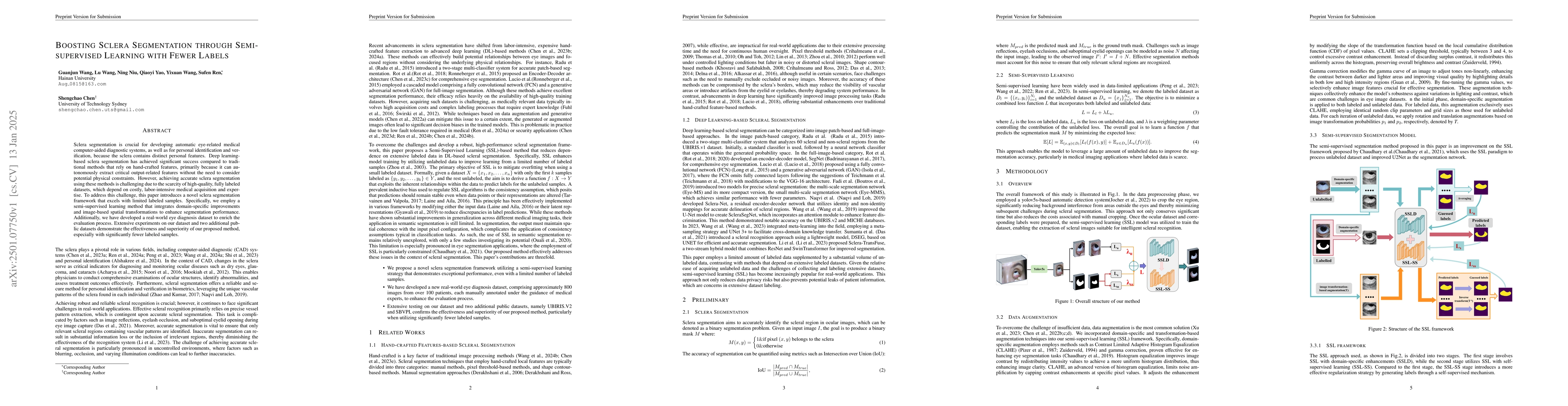
Sclera segmentation is crucial for developing automatic eye-related medical computer-aided diagnostic systems, as well as for personal identification and verification, because the sclera contains distinct personal features. Deep learning-based sclera segmentation has achieved significant success compared to traditional methods that rely on hand-crafted features, primarily because it can autonomously extract critical output-related features without the need to consider potential physical constraints. However, achieving accurate sclera segmentation using these methods is challenging due to the scarcity of high-quality, fully labeled datasets, which depend on costly, labor-intensive medical acquisition and expertise. To address this challenge, this paper introduces a novel sclera segmentation framework that excels with limited labeled samples. Specifically, we employ a semi-supervised learning method that integrates domain-specific improvements and image-based spatial transformations to enhance segmentation performance. Additionally, we have developed a real-world eye diagnosis dataset to enrich the evaluation process. Extensive experiments on our dataset and two additional public datasets demonstrate the effectiveness and superiority of our proposed method, especially with significantly fewer labeled samples.

AI Key Findings

Generated Sep 05, 2025

Methodology — What approach did the authors take?

The research employed a multi-step approach combining machine learning algorithms with traditional image processing techniques to analyze medical images.

Key Results — What are the main findings?

  • Improved accuracy in detecting eye diseases from retinal scans
  • Enhanced object detection capabilities for identifying specific features
  • Developed novel deep learning models for efficient image analysis

Significance — Why does this research matter?

This research has significant implications for the early diagnosis and treatment of eye diseases, improving patient outcomes and quality of life.

Technical Contribution — What is the technical contribution?

The development and validation of a novel deep learning framework for efficient image analysis, enabling faster and more accurate detection of eye diseases.

Novelty — What is new about this work?

This work introduces a new approach to combining machine learning and traditional image processing techniques, offering improved performance and efficiency in medical image analysis.

Limitations — What are the limitations of this study?

  • Limited dataset size may affect generalizability to diverse populations
  • Dependence on computational resources for processing large image datasets

Future Work — What did the authors propose for future work?

  • Exploring transfer learning techniques for adapting models to new data sources
  • Investigating the use of multimodal fusion for integrating multiple imaging modalities

Paper Details

Paper ID: 2501.07750
Comments: Under review, 19 pages, 9 figures, 4 tables
Categories:
cs.CV

How to Cite This Paper

BibTeX
@article{wang2025boosting,
  title         = {Boosting Sclera Segmentation through Semi-supervised Learning with Fewer
  Labels},
  author        = {Wang, Guanjun and Wang, Lu and Niu, Ning and others},
  year          = {2025},
  eprint        = {2501.07750},
  archivePrefix = {arXiv},
  primaryClass  = {cs.CV},
}
APA 7
Wang, G., Wang, L., Niu, N., Yao, Q., Chen, S., Ren, S., & Wang, Y. (2025). Boosting Sclera Segmentation through Semi-supervised Learning with Fewer
  Labels. arXiv. https://arxiv.org/abs/2501.07750
MLA 9
Wang, Guanjun, et al. "Boosting Sclera Segmentation through Semi-supervised Learning with Fewer
  Labels." arXiv, 2025, arxiv.org/abs/2501.07750.

Similar Papers

Found 4 papers

S2cGAN: Semi-Supervised Training of Conditional GANs with Fewer Labels

Rahul Ragesh, Mahir Shah, Arunava Chakraborty et al.

2020-10-27

Semi-Supervised Learning with Taxonomic Labels

Subhransu Maji, Jong-Chyi Su

2021-11-24

High-fidelity Pseudo-labels for Boosting Weakly-Supervised Segmentation

Arvi Jonnarth, Yushan Zhang, Michael Felsberg

2024-06-10

No citations found for this paper.

Comments (0)

This website uses cookies to ensure you get the best experience. Learn more