Boosting transducer matrix sensitivity for 3D large field ultrasound localization microscopy using a multi-lens diffracting layer: a simulation study
Publication
Metrics
AI Quick Summary
This study simulates a new high-sensitivity 3D ultrasound localization microscopy approach using large diverging elements and an adapted beamforming technique to enhance sensitivity for whole brain imaging. The proposed method detected 93% of microbubbles in a synthetic phantom, indicating its potential for clinical translation in mapping cerebral microflows.
Paper Preview
Abstract
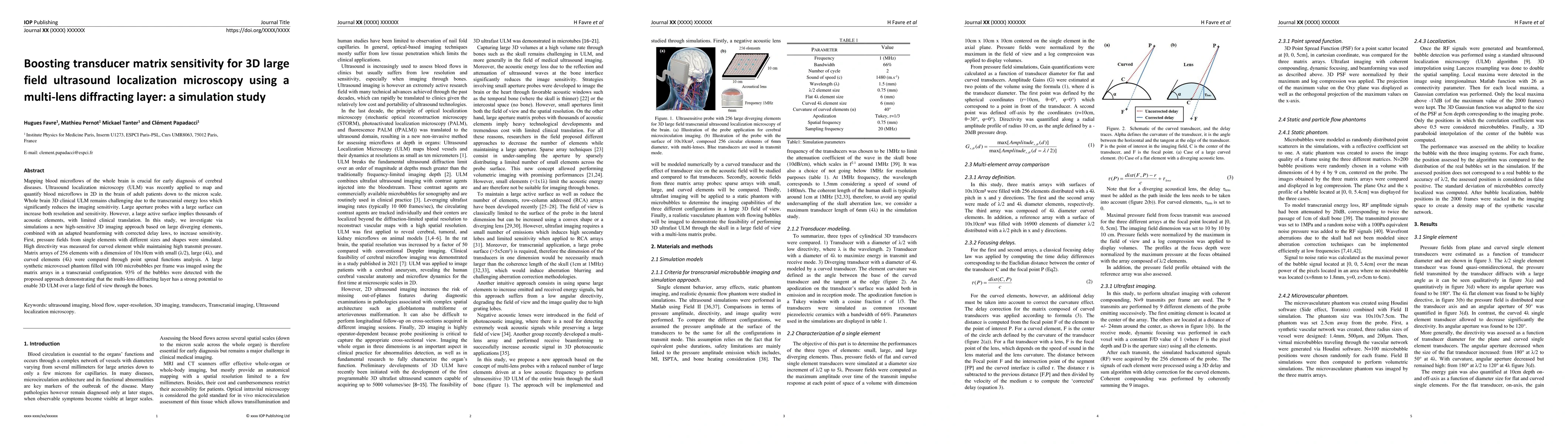
Mapping blood microflows of the whole brain is crucial for early diagnosis of cerebral diseases. Ultrasound localization microscopy (ULM) was recently applied to map and quantify blood microflows in 2D in the brain of adult patients down to the micron scale. Whole brain 3D clinical ULM remains challenging due to the transcranial energy loss which significantly reduces the imaging sensitivity. Large aperture probes with a large surface can increase both resolution and sensitivity. However, a large active surface implies thousands of acoustic elements, with limited clinical translation. In this study, we investigate via simulations a new high-sensitive 3D imaging approach based on large diverging elements, combined with an adapted beamforming with corrected delay laws, to increase sensitivity. First, pressure fields from single elements with different sizes and shapes were simulated. High directivity was measured for curved element while maintaining high transmit pressure. Matrix arrays of 256 elements with a dimension of 10x10 cm with small ( $\lambda$ /2), large (4 $\lambda$ ), and curved elements (4 $\lambda$ ) were compared through point spread functions analysis. A large synthetic microvessel phantom filled with 100 microbubbles per frame was imaged using the matrix arrays in a transcranial configuration. 93% of the bubbles were detected with the proposed approach demonstrating that the multi-lens diffracting layer has a strong potential to enable 3D ULM over a large field of view through the bones.
AI Key Findings
Get AI-generated insights about this paper's methodology, results, significance, and more — seven facets brought into focus.
Impact
Paper Details
Authors
PDF Preview
Key Terms
Citation Network
Current paper (gray), citations (green), references (blue)
Display is limited for performance on very large graphs.
Discussion 0