BoxNet introduces a novel weakly supervised deep learning approach for biomedical image segmentation using only box annotations, combining graph search and deep learning to generate fine object masks. The method achieves comparable accuracy to fully supervised methods with significantly reduced annotation effort and demonstrates superior performance across various segmentation tasks.
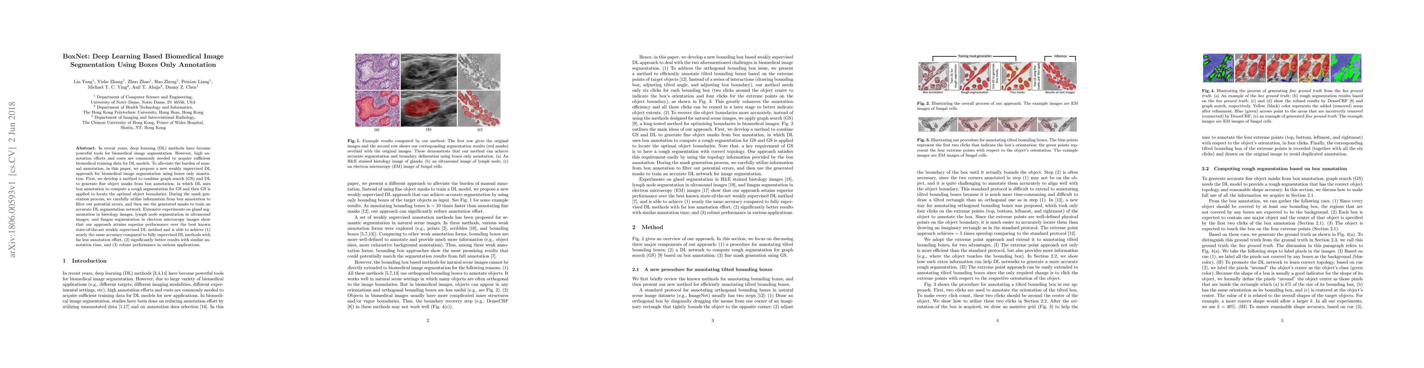
In recent years, deep learning (DL) methods have become powerful tools for biomedical image segmentation. However, high annotation efforts and costs are commonly needed to acquire sufficient biomedical training data for DL models. To alleviate the burden of manual annotation, in this paper, we propose a new weakly supervised DL approach for biomedical image segmentation using boxes only annotation. First, we develop a method to combine graph search (GS) and DL to generate fine object masks from box annotation, in which DL uses box annotation to compute a rough segmentation for GS and then GS is applied to locate the optimal object boundaries. During the mask generation process, we carefully utilize information from box annotation to filter out potential errors, and then use the generated masks to train an accurate DL segmentation network. Extensive experiments on gland segmentation in histology images, lymph node segmentation in ultrasound images, and fungus segmentation in electron microscopy images show that our approach attains superior performance over the best known state-of-the-art weakly supervised DL method and is able to achieve (1) nearly the same accuracy compared to fully supervised DL methods with far less annotation effort, (2) significantly better results with similar annotation time, and (3) robust performance in various applications.
This graph shows how this paper is connected to other research. The current paper is shown in dark gray, papers that cite it are in green, and papers it references are in blue. You can:
• Hover over any node to see the paper title
• Click a node to view that paper's details
• Drag nodes to rearrange the graph
• Use the zoom controls (bottom right) to zoom in/out
Title | Authors | Year | Actions |
---|---|---|---|
FrGNet: A fourier-guided weakly-supervised framework for nuclear instance segmentation
|
Peng Ling, Wenxiao Xiong
|
2025
|
View Paper |
GlandSAM: Injecting Morphology Knowledge Into Segment Anything Model for Label-Free Gland Segmentation
|
Qixiang Zhang, Yi Li, Cheng Xue, Haonan Wang, Xiaomeng Li
|
2024
|
View Paper |
Weakly supervised pre-training for brain tumor segmentation using principal axis measurements of tumor burden
|
Joshua E. Mckone, T. Lambrou, Xujiong Ye, James M. Brown
|
2024
|
View Paper |
Point annotations for nucleus segmentation in histopathology images via envelope enhancement network
|
Han Hong, Aiping Qu, Tongqing Xue
|
2024
|
View Paper |
Radiomics Analysis in Characterization of Salivary Gland Tumors on MRI: A Systematic Review
|
Kaijing Mao, L. M. Wong, Rongli Zhang, T. So, Zhiyi Shan, Kuo Feng Hung, Q. H. Ai
|
2023
|
View Paper |
The Contrastive Network With Convolution and Self-Attention Mechanisms for Unsupervised Cell Segmentation
|
Yuhang Zhao, Xianhao Shao, Cai Chen, Junlin Song, Chongxuan Tian, Wei Li
|
2023
|
View Paper |
Morphology-inspired Unsupervised Gland Segmentation via Selective Semantic Grouping
|
Qixiang Zhang, Yi Li, Cheng Xue, X. Li
|
2023
|
View Paper |
A review of open-source image analysis tools for mammalian cell culture: algorithms, features and implementations
|
Hafizi Malik, Ahmad Syahrin Idris, S. Toha, Izyan Mohd Idris, M. F. Daud, Nur Liyana Azmi
|
2023
|
View Paper |
Cyclic Learning: Bridging Image-Level Labels and Nuclei Instance Segmentation
|
Yang Zhou, Yongjian Wu, Z. Wang, Bingzheng Wei, Maode Lai, Jianzhong Shou, Yubo Fan, Yan Xu
|
2023
|
View Paper |
Weakly Supervised Cell-Instance Segmentation with Two Types of Weak Labels by Single Instance Pasting
|
Kazuya Nishimura, Ryoma Bise
|
2023
|
View Paper |
Coarse Retinal Lesion Annotations Refinement via Prototypical Learning
|
Qinji Yu, K. Dang, Ziyu Zhou, Y. Chen, Xiucai Ding
|
2022
|
View Paper |
Learning to Segment Fine Structures Under Image-Level Supervision With an Application to Nematode Segmentation*
|
Long Chen, M. Strauch, M. Daub, Hans-Georg Luigs, Marcus Jansen, D. Merhof
|
2022
|
View Paper |
Microscopy analysis neural network to solve detection, enumeration and segmentation from image-level annotations
|
Anthony Bilodeau, Constantin V. L. Delmas, M. Parent, P. De Koninck, A. Durand, Flavie Lavoie-Cardinal
|
2022
|
View Paper |
CST: A Multitask Learning Framework for Colorectal Cancer Region Mining Based on Transformer
|
D. Sui, Kang Zhang, Weifeng Liu, Jing Chen, Xiaoxuan Ma, Z. Tian
|
2021
|
View Paper |
PiPo-Net: A Semi-automatic and Polygon-based Annotation Method for Pathological Images
|
Yuqi Fang, Delong Zhu, Niyun Zhou, Li Liu, Jianhua Yao
|
2021
|
View Paper |
A survey on applications of deep learning in microscopy image analysis
|
Zhichao Liu, Luhong Jin, Jincheng Chen, Qiuyu Fang, S. Ablameyko, Zhaozheng Yin, Yingke Xu
|
2021
|
View Paper |
MICRA-Net: MICRoscopy Analysis Neural Network to solve detection, classification, and segmentation from a single simple auxiliary task
|
Anthony Bilodeau, Constantin V. L. Delmas, M. Parent, P. De Koninck, A. Durand, Flavie Lavoie-Cardinal
|
2020
|
View Paper |
TissueWand, a Rapid Histopathology Annotation Tool
|
Martin Lindvall, Alexander Sanner, F. Petre, K. Lindman, D. Treanor, C. Lundström, J. Löwgren
|
2020
|
View Paper |
Weakly Supervised Deep Nuclei Segmentation Using Partial Points Annotation in Histopathology Images
|
Hui Qu, Pengxiang Wu, Qiaoying Huang, Jingru Yi, Zhennan Yan, Kang Li, G. Riedlinger, S. De, Shaoting Zhang, Dimitris N. Metaxas
|
2020
|
View Paper |
Train, Learn, Expand, Repeat
|
Abhijeet Parida, Aadhithya Sankar, R. Eisawy, Tom Finck, B. Wiestler, F. Pfister, Julia Moosbauer
|
2020
|
View Paper |
An Annotation Sparsification Strategy for 3D Medical Image Segmentation via Representative Selection and Self-Training
|
Hao Zheng, Yizhe Zhang, Lin Yang, Chaoli Wang, D. Chen
|
2020
|
View Paper |
Deep neural network models for computational histopathology: A survey
|
C. Srinidhi, Ozan Ciga, Anne L. Martel
|
2019
|
View Paper |
Learning to segment images with classification labels
|
Ozan Ciga, Anne L. Martel
|
2019
|
View Paper |
A Survey on Deep Learning of Small Sample in Biomedical Image Analysis
|
Pengyi Zhang, Yunxin Zhong, Yulin Deng, Xiaoying Tang, Xiaoqiong Li
|
2019
|
View Paper |
Biomedical Image Segmentation via Representative Annotation
|
Hao Zheng, Lin Yang, Jianxu Chen, Jun Han, Yizhe Zhang, Peixian Liang, Zhuo Zhao, Chaoli Wang, D. Chen
|
2019
|
View Paper |
Weakly Supervised Deep Nuclei Segmentation using Points Annotation in Histopathology Images
|
Hui Qu, Pengxiang Wu, Qiaoying Huang, Jingru Yi, G. Riedlinger, S. De, Dimitris N. Metaxas
|
2019
|
View Paper |
DRPnet - Automated Particle Picking in Cryo-Electron Micrographs using Deep Regression
|
Nguyen P. Nguyen, Jacob Gotberg, I. Ersoy, F. Bunyak, Tommi White
|
2019
|
View Paper |
Review of Histopathological Image Segmentation via current Deep Learning Approaches
|
Manju Dabass, R. Vig, Sharda Vashisth
|
2018
|
View Paper |
Considerations for data acquisition and modeling strategies: Mitosis detection in computational pathology
|
Zongliang Ji, Philip Rosenfield, Christina Eng, Sarah Bettigole, Danielle C. Gibson, Hamid Masoudi, Matthew Hanna, Nicolò Fusi, Kristen Severson
|
2023
|
View Paper |
Multi-modal Cell Segmentation based on U-Net++ and Attention Gate
|
Xinye Yang, Hao Chen
|
2022
|
View Paper |
Medical Image Analysis
|
Peng Ling
|
View Paper |
Published: 2018-07-02
Published: 2017-06-16
Published: 2020-03-16
Shaoting Zhang, Xiangde Luo, Guotai Wang, Kang Li, Shuojue Yang, Yijie Qu, Ran Gu, Shuwei Zhai, Qianfei Zhao
Published: 2023-02-14
Comments (0)