Brain Stroke Segmentation Using Deep Learning Models: A Comparative Study
Publication
Metrics
AI Quick Summary
This study compares four deep learning models—Transformer-based DAE-Former, CNN-based LKA and DLKA, hybrid FCT, and nnUNet—for brain stroke segmentation, finding nnUNet best due to its robust design and effective preprocessing/postprocessing, while highlighting Transformers' limitations in handling dataset variabilities.
Paper Preview
Abstract
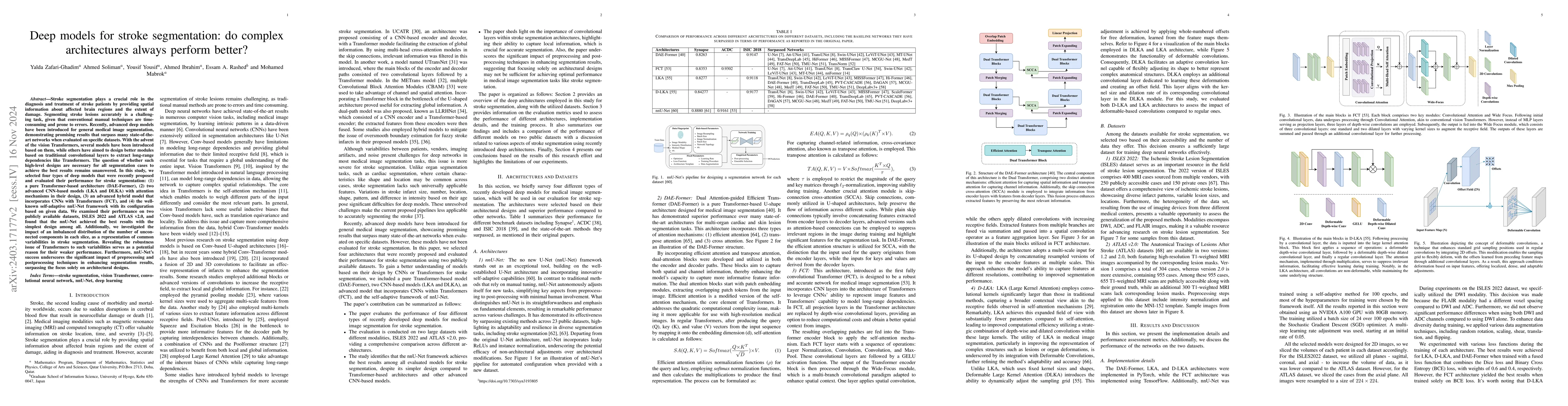
Stroke segmentation plays a crucial role in the diagnosis and treatment of stroke patients by providing spatial information about affected brain regions and the extent of damage. Segmenting stroke lesions accurately is a challenging task, given that conventional manual techniques are time consuming and prone to errors. Recently, advanced deep models have been introduced for general medical image segmentation, demonstrating promising results that surpass many state of the art networks when evaluated on specific datasets. With the advent of the vision Transformers, several models have been introduced based on them, while others have aimed to design better modules based on traditional convolutional layers to extract long-range dependencies like Transformers. The question of whether such high-level designs are necessary for all segmentation cases to achieve the best results remains unanswered. In this study, we selected four types of deep models that were recently proposed and evaluated their performance for stroke segmentation: a pure Transformer-based architecture (DAE-Former), two advanced CNN-based models (LKA and DLKA) with attention mechanisms in their design, an advanced hybrid model that incorporates CNNs with Transformers (FCT), and the well-known self-adaptive nnUNet framework with its configuration based on given data. We examined their performance on two publicly available datasets, and found that the nnUNet achieved the best results with the simplest design among all. Revealing the robustness issue of Transformers to such variabilities serves as a potential reason for their weaker performance. Furthermore, nnUNet's success underscores the significant impact of preprocessing and postprocessing techniques in enhancing segmentation results, surpassing the focus solely on architectural designs
AI Key Findings
Get AI-generated insights about this paper's methodology, results, significance, and more — seven facets brought into focus.
Impact
Paper Details
Authors
PDF Preview
Key Terms
Citation Network
Current paper (gray), citations (green), references (blue)
Display is limited for performance on very large graphs.
Discussion 0