This paper explores the use of NeuroNet, incorporating ResNet and FCN, for automatic segmentation of brain tissues in MRI images. Various pre-processing techniques and hyper-parameter tuning were employed to enhance model performance, achieving a Dice Similarity Coefficient of 0.84 for CSF, and 0.94 for both GM and WM, indicating significant improvements through pre-processing.
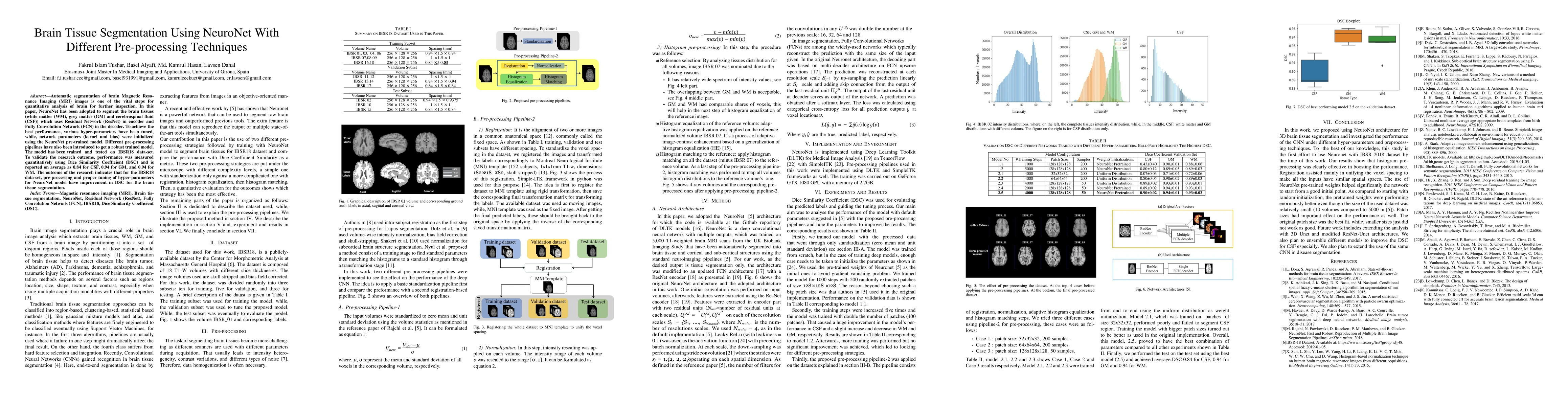
Automatic segmentation of brain Magnetic Resonance Imaging (MRI) images is one of the vital steps for quantitative analysis of brain for further inspection. In this paper, NeuroNet has been adopted to segment the brain tissues (white matter (WM), grey matter (GM) and cerebrospinal fluid (CSF)) which uses Residual Network (ResNet) in encoder and Fully Convolution Network (FCN) in the decoder. To achieve the best performance, various hyper-parameters have been tuned, while, network parameters (kernel and bias) were initialized using the NeuroNet pre-trained model. Different pre-processing pipelines have also been introduced to get a robust trained model. The model has been trained and tested on IBSR18 data-set. To validate the research outcome, performance was measured quantitatively using Dice Similarity Coefficient (DSC) and is reported on average as 0.84 for CSF, 0.94 for GM, and 0.94 for WM. The outcome of the research indicates that for the IBSR18 data-set, pre-processing and proper tuning of hyper-parameters for NeuroNet model have improvement in DSC for the brain tissue segmentation.
Get AI-generated insights about this paper's methodology, results, and significance.
Current paper (gray), citations (green), references (blue)
Mohammad Imran Hossain, Muhammad Zain Amin, Daniel Tweneboah Anyimadu et al.
Ye Tian, Jia Guo, Xuzhe Zhang et al.
Tom Vercauteren, Lucas Fidon, Ali Gholipour et al.
Title | Authors | Year | Actions |
---|---|---|---|
A Systematic Study of Artificial Intelligence-Based Methods for Detecting Brain Tumors
|
Sanjeet Kumar, Urmila Pilania, Neha Nandal
|
2023
|
View Paper |
A survey, review, and future trends of skin lesion segmentation and classification
|
M. Hasan, Md. Asif Ahamad, C. Yap, Guang Yang
|
2022
|
View Paper |
Ensemble of Convolutional Neural Networks to diagnose Acute Lymphoblastic Leukemia from microscopic images
|
Chayan Mondal, Md. Kamrul Hasan, Mohiudding Ahmad, M. Awal, Md. Tasnim Jawad, Aishwariya Dutta, Md. Rabiul Islam, M. Moni
|
2021
|
View Paper |
Automated multiclass tissue segmentation of clinical brain MRIs with lesions
|
D. Weiss, Rachit Saluja, L. Xie, J. Gee, L. Sugrue, Abhijeet Pradhan, R., N. Bryan, A. Rauschecker, J. Rudie
|
2021
|
View Paper |
Acute Lymphoblastic Leukemia Detection from Microscopic Images Using Weighted Ensemble of Convolutional Neural Networks
|
Chayan Mondal, Md. Kamrul Hasan, Md. Tasnim Jawad, Aishwariya Dutta, M. Islam, M. Awal, Mohiudding Ahmad
|
2021
|
View Paper |
COVID-19 identification from volumetric chest CT scans using a progressively resized 3D-CNN incorporating segmentation, augmentation, and class-rebalancing
|
M. K. Hasan, Md. Tasnim Jawad, Kazi N. Hasan, Sajal Basak Partha, Md. Masum Al Masba
|
2021
|
View Paper |
DermoExpert: Skin lesion classification using a hybrid convolutional neural network through segmentation, transfer learning, and augmentation
|
M. Hasan, M. E. Elahi, Md. Ashraful Alam, Md. Tasnim Jawad, R. Martí
|
2021
|
View Paper |
VOLUMETRIC AND VBM MEASURES IN 1.5-5 YEARS OLD CHILDREN OBTAINED USING ADULT SEGMENTATION TOOLS
|
Elena Ukharova
|
2020
|
View Paper |
Convolutional neural network MRI segmentation for fast and robust optimization of transcranial electrical current stimulation of the human brain
|
Carla Sendra-Balcells, R. Salvador, J. Pedro, M. Biagi, Charlène Aubinet, B. Manor, A. Thibaut, S. Laureys, K. Lekadir, G. Ruffini
|
2020
|
View Paper |
Skin Lesion Analysis: A State-of-the-Art Survey, Systematic Review, and Future Trends
|
M. Hasan, Md. Asif Ahamad, C. Yap, Guang Yang
|
2022
|
View Paper |
Incorporating global multiplicative decomposition and local statistical information for brain tissue segmentation and bias field estimation
|
Haiping Xu, Geng Lin
|
2021
|
View Paper |
Comments (0)