Brain Tissues Segmentation on MR Perfusion Images Using CUSUM Filter for Boundary Pixels
Publication
Metrics
AI Quick Summary
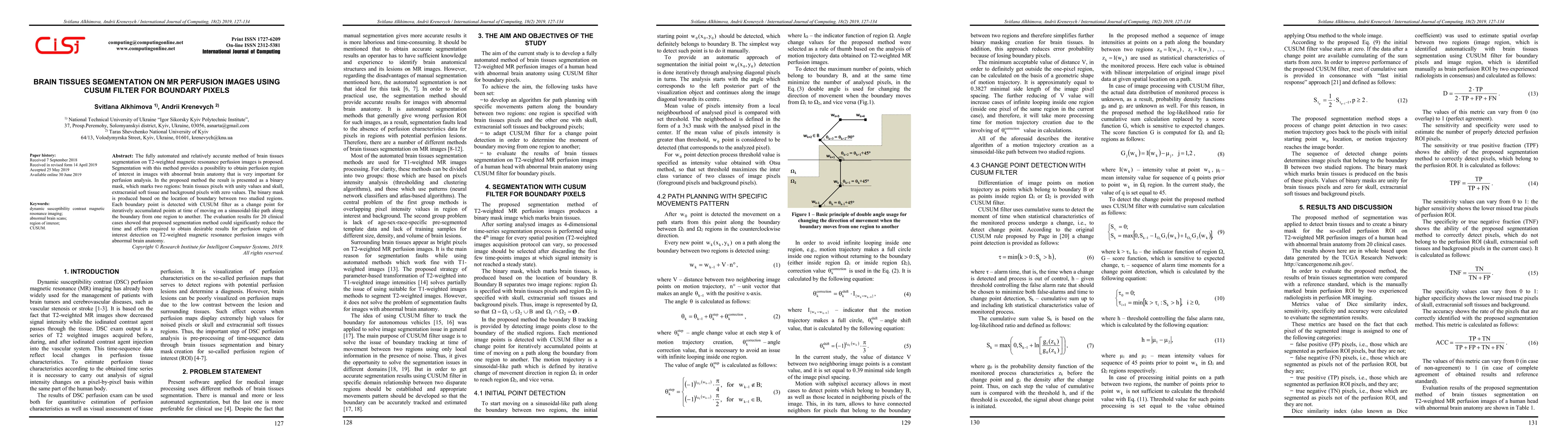
The paper proposes a fully automated method for brain tissues segmentation on T2-weighted MR perfusion images using a CUSUM filter to detect boundary pixels, yielding a binary mask for accurate perfusion region of interest detection, significantly reducing segmentation time and effort. Clinical evaluation on 20 cases confirmed its effectiveness.
Paper Preview
Abstract
The fully automated and relatively accurate method of brain tissues segmentation on T2-weighted magnetic resonance perfusion images is proposed. Segmentation with this method provides a possibility to obtain perfusion region of interest on images with abnormal brain anatomy that is very important for perfusion analysis. In the proposed method the result is presented as a binary mask, which marks two regions: brain tissues pixels with unity values and skull, extracranial soft tissue and background pixels with zero values. The binary mask is produced based on the location of boundary between two studied regions. Each boundary point is detected with CUSUM filter as a change point for iteratively accumulated points at time of moving on a sinusoidal-like path along the boundary from one region to another. The evaluation results for 20 clinical cases showed that proposed segmentation method could significantly reduce the time and efforts required to obtain desirable results for perfusion region of interest detection on T2-weighted magnetic resonance perfusion images with abnormal brain anatomy.
AI Key Findings
Get AI-generated insights about this paper's methodology, results, significance, and more — seven facets brought into focus.
Impact
Paper Details
Authors
PDF Preview
Key Terms
Citation Network
Current paper (gray), citations (green), references (blue)
Display is limited for performance on very large graphs.
Discussion 0