Brain Tumor Segmentation in MRI Images with 3D U-Net and Contextual Transformer
Publication
Metrics
AI Quick Summary
This paper proposes a 3D U-Net model integrated with a Context Transformer (CoT) for precise brain tumor segmentation in MRI images, achieving high Dice scores of 82.0%, 81.5%, and 89.0% for Enhancing Tumor, Tumor Core, and Whole Tumor respectively, outperforming existing methods.
Paper Preview
Abstract
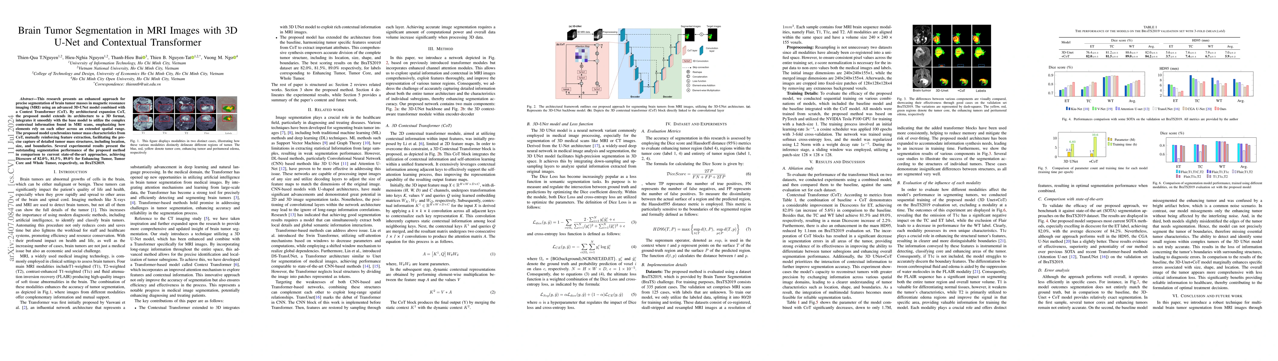
This research presents an enhanced approach for precise segmentation of brain tumor masses in magnetic resonance imaging (MRI) using an advanced 3D-UNet model combined with a Context Transformer (CoT). By architectural expansion CoT, the proposed model extends its architecture to a 3D format, integrates it smoothly with the base model to utilize the complex contextual information found in MRI scans, emphasizing how elements rely on each other across an extended spatial range. The proposed model synchronizes tumor mass characteristics from CoT, mutually reinforcing feature extraction, facilitating the precise capture of detailed tumor mass structures, including location, size, and boundaries. Several experimental results present the outstanding segmentation performance of the proposed method in comparison to current state-of-the-art approaches, achieving Dice score of 82.0%, 81.5%, 89.0% for Enhancing Tumor, Tumor Core and Whole Tumor, respectively, on BraTS2019.
AI Key Findings
Get AI-generated insights about this paper's methodology, results, significance, and more — seven facets brought into focus.
Impact
Authors
PDF Preview
Key Terms
Citation Network
Current paper (gray), citations (green), references (blue)
Display is limited for performance on very large graphs.
Discussion 0