Brain Tumor Segmentation using Enhanced U-Net Model with Empirical Analysis
Publication
Metrics
AI Quick Summary
This paper proposes an enhanced U-Net model for segmenting brain tumors in MRI images, trained on the BraTS dataset. The improved model effectively distinguishes necrotic, edematous, growing, and healthy tissues, achieving high Dice scores of 0.8717, 0.9506, and 0.9427 respectively, demonstrating its efficacy in clinical applications.
Paper Preview
Abstract
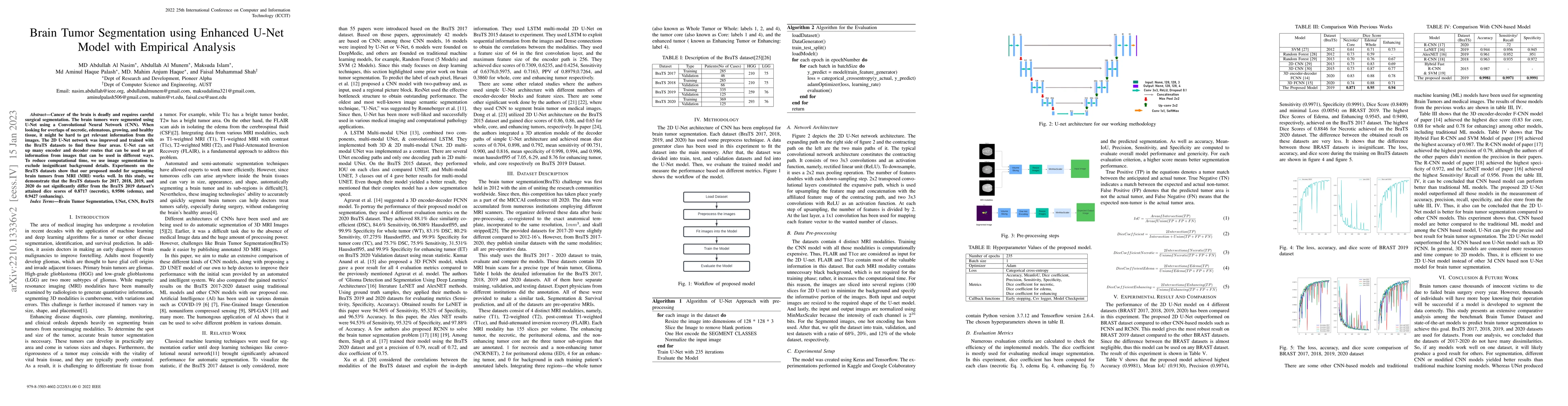
Cancer of the brain is deadly and requires careful surgical segmentation. The brain tumors were segmented using U-Net using a Convolutional Neural Network (CNN). When looking for overlaps of necrotic, edematous, growing, and healthy tissue, it might be hard to get relevant information from the images. The 2D U-Net network was improved and trained with the BraTS datasets to find these four areas. U-Net can set up many encoder and decoder routes that can be used to get information from images that can be used in different ways. To reduce computational time, we use image segmentation to exclude insignificant background details. Experiments on the BraTS datasets show that our proposed model for segmenting brain tumors from MRI (MRI) works well. In this study, we demonstrate that the BraTS datasets for 2017, 2018, 2019, and 2020 do not significantly differ from the BraTS 2019 dataset's attained dice scores of 0.8717 (necrotic), 0.9506 (edema), and 0.9427 (enhancing).
AI Key Findings
Get AI-generated insights about this paper's methodology, results, significance, and more — seven facets brought into focus.
Impact
Paper Details
Authors
PDF Preview
Key Terms
Citation Network
Current paper (gray), citations (green), references (blue)
Display is limited for performance on very large graphs.
Discussion 0