Publication

Published: Feb 25, 2026
Categories: cs.CV

Metrics

Source: ArXiv

Quick Actions

View PDF

Quick Answers

What methodology did the authors use?

Weight-inflated 2D vision encoder is converted to native 3D, followed by staged alignment—contrastive grounding, projector warmup, and LoRA-based linguistic specialization—using a soft‑prompted MedGemma LLM. More in Methodology →

What are the key results?

Clinical Pathology F1 of 0.951, a 130% gain over a 2D baseline — Perfect specificity on healthy controls More in Key Results →

Why is this work significant?

The framework delivers clinically accurate, structured brain‑MRI reports while preserving volumetric context, reducing hallucinations and enabling reliable neuroradiological decision support. More in Significance →

What are the main limitations?

Lateralization errors (~15 %) and occasional diffuse‑glioma under‑reporting — Model trained only on FLAIR, limiting generalizability to other sequences More in Limitations →

Paper Preview

Abstract

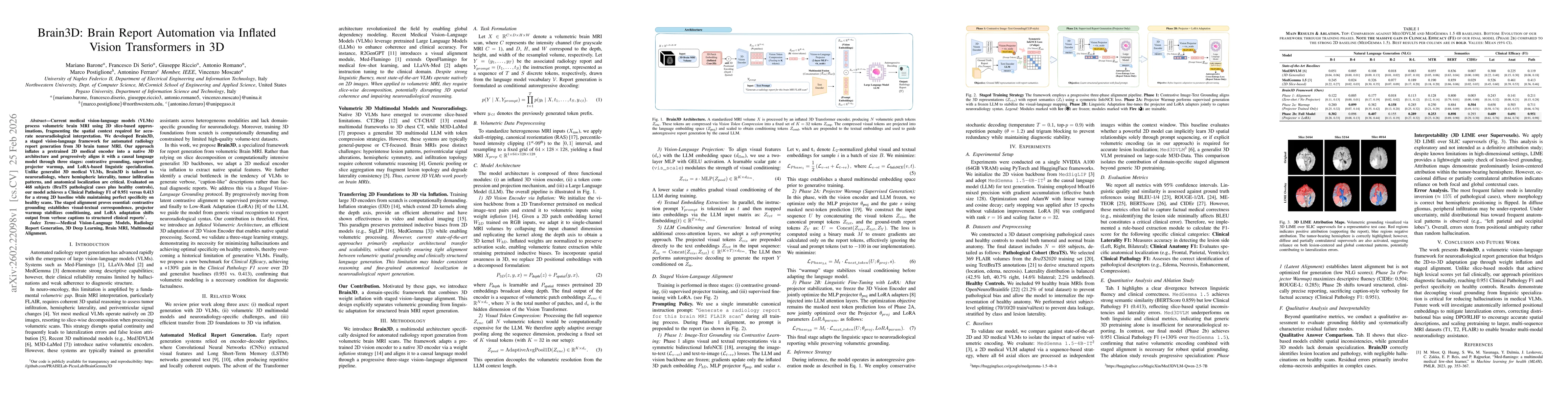
Current medical vision-language models (VLMs) process volumetric brain MRI using 2D slice-based approximations, fragmenting the spatial context required for accurate neuroradiological interpretation. We developed \textbf{Brain3D}, a staged vision-language framework for automated radiology report generation from 3D brain tumor MRI. Our approach inflates a pretrained 2D medical encoder into a native 3D architecture and progressively aligns it with a causal language model through three stages: contrastive grounding, supervised projector warmup, and LoRA-based linguistic specialization. Unlike generalist 3D medical VLMs, \textbf{Brain3D} is tailored to neuroradiology, where hemispheric laterality, tumor infiltration patterns, and anatomical localization are critical. Evaluated on 468 subjects (BraTS pathological cases plus healthy controls), our model achieves a Clinical Pathology F1 of 0.951 versus 0.413 for a strong 2D baseline while maintaining perfect specificity on healthy scans. The staged alignment proves essential: contrastive grounding establishes visual-textual correspondence, projector warmup stabilizes conditioning, and LoRA adaptation shifts output from verbose captions to structured clinical reports\footnote{Our code is publicly available for transparency and reproducibility

AI Key Findings

Generated Feb 26, 2026

Methodology — What approach did the authors take?

Weight-inflated 2D vision encoder is converted to native 3D, followed by staged alignment—contrastive grounding, projector warmup, and LoRA-based linguistic specialization—using a soft‑prompted MedGemma LLM.

Key Results — What are the main findings?

  • Clinical Pathology F1 of 0.951, a 130% gain over a 2D baseline
  • Perfect specificity on healthy controls
  • Ablation shows staged alignment is essential, with Phase 2b delivering the best performance

Significance — Why does this research matter?

The framework delivers clinically accurate, structured brain‑MRI reports while preserving volumetric context, reducing hallucinations and enabling reliable neuroradiological decision support.

Technical Contribution — What is the technical contribution?

A staged vision‑language alignment pipeline that inflates a 2D encoder to 3D, compresses visual tokens, and progressively aligns with a causal LLM via contrastive grounding, projector warmup, and LoRA adaptation.

Novelty — What is new about this work?

First domain‑specific 3D VLM for neuroradiology that combines weight inflation with progressive alignment, achieving superior clinical factuality over generic 3D VLMs.

Limitations — What are the limitations of this study?

  • Lateralization errors (~15 %) and occasional diffuse‑glioma under‑reporting
  • Model trained only on FLAIR, limiting generalizability to other sequences

Future Work — What did the authors propose for future work?

  • Incorporate anatomically informed positional embeddings and RLHF/DPO to improve spatial precision
  • Scale pretraining to multi‑sequence MRI (T1, T2, FLAIR) for broader clinical applicability

Paper Details

Paper ID: 2602.22098
Categories:
cs.CV

How to Cite This Paper

BibTeX
@article{ferraro2026braind,
  title         = {Brain3D: Brain Report Automation via Inflated Vision Transformers in 3D},
  author        = {Ferraro, Antonino and Romano, Antonio and Serio, Francesco Di and others},
  year          = {2026},
  eprint        = {2602.22098},
  archivePrefix = {arXiv},
  primaryClass  = {cs.CV},
}
APA 7
Ferraro, A., Romano, A., Serio, F., Riccio, G., Postiglione, M., Barone, M., & Moscato, V. (2026). Brain3D: Brain Report Automation via Inflated Vision Transformers in 3D. arXiv. https://arxiv.org/abs/2602.22098
MLA 9
Ferraro, Antonino, et al. "Brain3D: Brain Report Automation via Inflated Vision Transformers in 3D." arXiv, 2026, arxiv.org/abs/2602.22098.

Similar Papers

Found 4 papers

Brain3D: EEG-to-3D Decoding of Visual Representations via Multimodal Reasoning

Emanuele Balloni, Emanuele Frontoni, Chiara Matti et al.

2026-04-09

3D Vision with Transformers: A Survey

Jiale Cao, Rao Muhammad Anwer, Salman Khan et al.

2022-08-09

Brain-Inspired Stepwise Patch Merging for Vision Transformers

Yonghao Yu, Dongcheng Zhao, Guobin Shen et al.

2024-09-11

Comments (0)

This website uses cookies to ensure you get the best experience. Learn more