Breast Cancer Classification in Deep Ultraviolet Fluorescence Images Using a Patch-Level Vision Transformer Framework

Source: ArXiv

AI Quick Summary

A new deep learning framework using patch-level vision transformer model improves breast cancer classification accuracy to 98.33% on DUV fluorescence images, outperforming conventional methods.

Share: Share on X LinkedIn

Summary

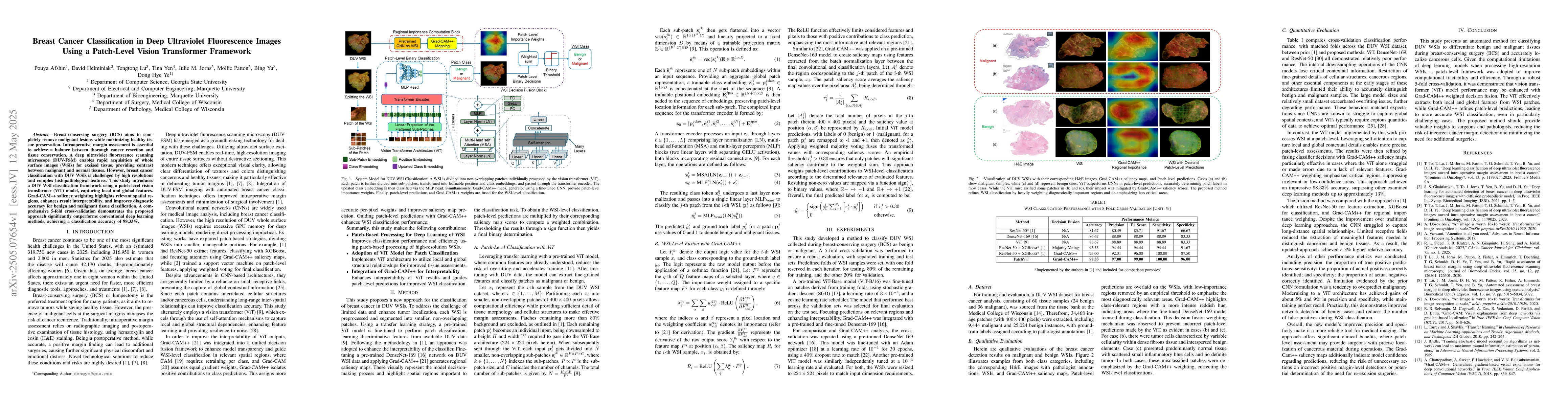
Breast-conserving surgery (BCS) aims to completely remove malignant lesions while maximizing healthy tissue preservation. Intraoperative margin assessment is essential to achieve a balance between thorough cancer resection and tissue conservation. A deep ultraviolet fluorescence scanning microscope (DUV-FSM) enables rapid acquisition of whole surface images (WSIs) for excised tissue, providing contrast between malignant and normal tissues. However, breast cancer classification with DUV WSIs is challenged by high resolutions and complex histopathological features. This study introduces a DUV WSI classification framework using a patch-level vision transformer (ViT) model, capturing local and global features. Grad-CAM++ saliency weighting highlights relevant spatial regions, enhances result interpretability, and improves diagnostic accuracy for benign and malignant tissue classification. A comprehensive 5-fold cross-validation demonstrates the proposed approach significantly outperforms conventional deep learning methods, achieving a classification accuracy of 98.33%.

AI Key Findings

Generated Jun 08, 2025

Methodology

The research employs a patch-level vision transformer (ViT) model for classifying deep ultraviolet fluorescence images (DUV WSIs) of excised breast tissue, utilizing Grad-CAM++ for saliency weighting and enhancing interpretability.

Key Results

  • The proposed approach achieves a classification accuracy of 98.33% in 5-fold cross-validation, significantly outperforming conventional deep learning methods.
  • Grad-CAM++ saliency weighting highlights relevant spatial regions, improving diagnostic accuracy for benign and malignant tissue classification.

Significance

This research is important for intraoperative margin assessment in breast-conserving surgery, as it enables rapid and accurate classification of malignant lesions using DUV WSIs, potentially improving cancer resection and tissue conservation balance.

Technical Contribution

The patch-level ViT framework with Grad-CAM++ saliency weighting for DUV WSI classification, enhancing both accuracy and interpretability.

Novelty

This work introduces a novel application of vision transformers at the patch level for DUV WSI classification, outperforming existing deep learning methods with improved diagnostic accuracy and result interpretability.

Limitations

  • The study did not address the generalizability of the model to diverse patient populations or imaging equipment.
  • No information was provided on computational resources required for training and inference.

Future Work

  • Investigate the model's performance on diverse patient cohorts and imaging devices.
  • Explore methods to reduce computational requirements for practical implementation.

Paper Details

PDF Preview

Download PDF

No citations found for this paper.

Comments (0)

This website uses cookies to ensure you get the best experience. Learn more