Quick Summary:

This study uses an AI-based algorithm to quantify breast density in MRI, finding consistent density values across datasets and a correlation with mammographic density, while also highlighting unique density components captured only by MRI. Future research aims to integrate MR density assessments to enhance breast cancer risk prediction.

Summary

Mammographic breast density is a well-established risk factor for breast cancer. Recently there has been interest in breast MRI as an adjunct to mammography, as this modality provides an orthogonal and highly quantitative assessment of breast tissue. However, its 3D nature poses analytic challenges related to delineating and aggregating complex structures across slices. Here, we applied an in-house machine-learning algorithm to assess breast density on normal breasts in three MRI datasets. Breast density was consistent across different datasets (0.104 - 0.114). Analysis across different age groups also demonstrated strong consistency across datasets and confirmed a trend of decreasing density with age as reported in previous studies. MR breast density was correlated with mammographic breast density, although some notable differences suggest that certain breast density components are captured only on MRI. Future work will determine how to integrate MR breast density with current tools to improve future breast cancer risk prediction.

AI Key Findings

Generated Jun 09, 2025

Methodology

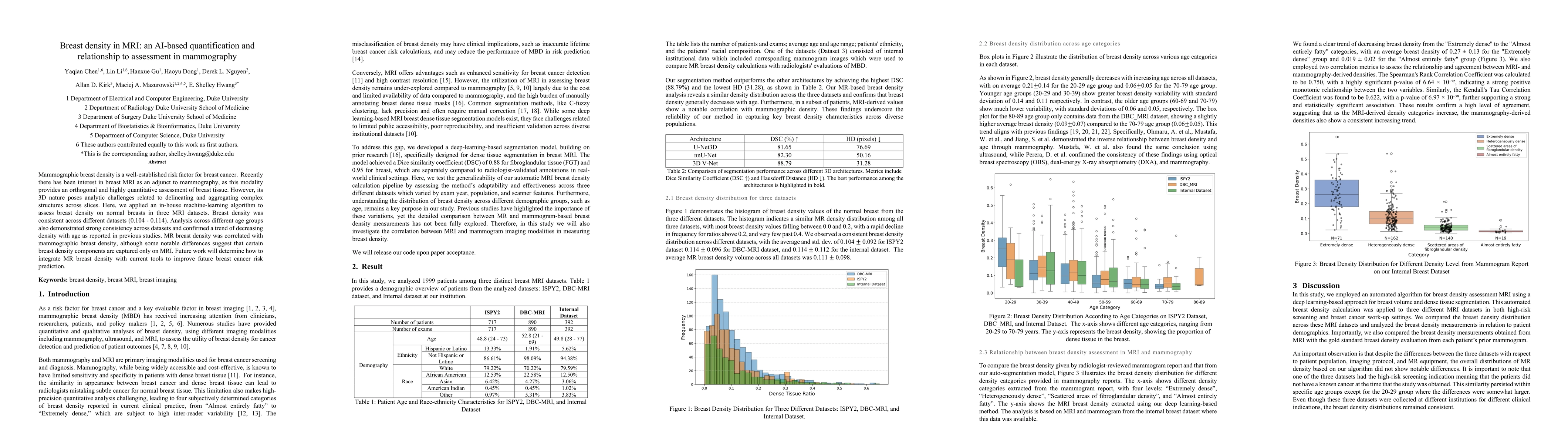
This study employed an in-house machine-learning algorithm to assess breast density on normal breasts using three MRI datasets. The algorithm utilized deep learning-based breast volume and dense tissue segmentation, comparing breast density distribution across datasets and analyzing measurements in relation to patient demographics. The study also compared MRI-derived breast density with mammography-based gold standard evaluations.

Key Results

  • Breast density was consistent across different MRI datasets (0.104 - 0.114).
  • Analysis across different age groups confirmed a trend of decreasing density with age.
  • MRI breast density was correlated with mammographic breast density, although some differences suggest that certain density components are captured only on MRI.
  • MRI-based breast density evaluation showed promising advantages in segmentation accuracy and BI-RADS classification performance compared to existing mammography-based approaches.
  • The model achieved a Dice Coefficient of 88% for dense tissue segmentation and 95% for overall breast segmentation.

Significance

This research demonstrates the reliability of MRI in assessing breast density, which can help identify distinct subgroups within high-risk categories and improve breast cancer risk prediction models. The study also highlights the potential of MRI to provide more accurate volumetric measurements of dense tissue compared to mammography.

Technical Contribution

The development and application of a deep learning-based algorithm for breast density assessment using MRI, demonstrating high consistency and accuracy across diverse datasets.

Novelty

This work presents a novel approach to breast density quantification using MRI, offering improved accuracy and potential for identifying distinct subgroups within high-risk categories, which sets it apart from existing mammography-based methods.

Limitations

  • The study's patient population undergoing breast MRI are typically high-risk screening patients or those with known breast cancer, which may limit the generalizability of results to the general screening population.
  • The model lacks a quantitative measure of the spatial distribution of dense tissue, which could influence risk assessment outcomes.

Future Work

  • Investigate how to integrate MR breast density with current tools to improve future breast cancer risk prediction.
  • Explore settings where MR may provide additional, orthogonal data to mammography assessments.

Paper Details

PDF Preview

Download PDF

Key Terms

No key terms available for this paper.

Citation Network

Current paper (gray), citations (green), references (blue)

Current Paper
Citations
References
Click to view

No citations found for this paper.

Comments (0)

This website uses cookies to ensure you get the best experience. Learn more