ArxivLens

Quick Summary:

This paper demonstrates the use of focused ion beam-scanning electron microscopy (FIB-SEM) tomography for high-resolution characterization of large colloidal assemblies, providing detailed insights into particle positions and orientations. This method enables the study of assemblies beyond the reach of conventional microscopy, crucial for understanding the collective properties of nanoparticle materials.

Journal Reference: Nanoscale, 2019, 11, 5304-5316
DOI: 10.1039/C8NR09753D
arXiv Id: 1903.09989
Comments: 17 pages, 4 figures, Supplemental Information at articles webpage: https://doi.org/10.1039/C8NR09753D
Date Published: 2019-03-27
Date Updated: 2019-03-27

Key Terms (TF-IDF):

tomography (0.263)
microscopy (0.244)
containing (0.210)
particle (0.189)
sized (0.185)
electron (0.178)
disordered (0.176)
conventional (0.174)
ordered (0.165)
collective (0.164)
structural (0.153)
transmission (0.152)
characterization (0.149)
individual (0.136)
small (0.128)
resolution (0.125)
micron (0.119)
gold (0.118)
nanoparticles (0.117)
recently developed (0.116)
License: http://arxiv.org/licenses/nonexclusive-distrib/1.0/

Summary:

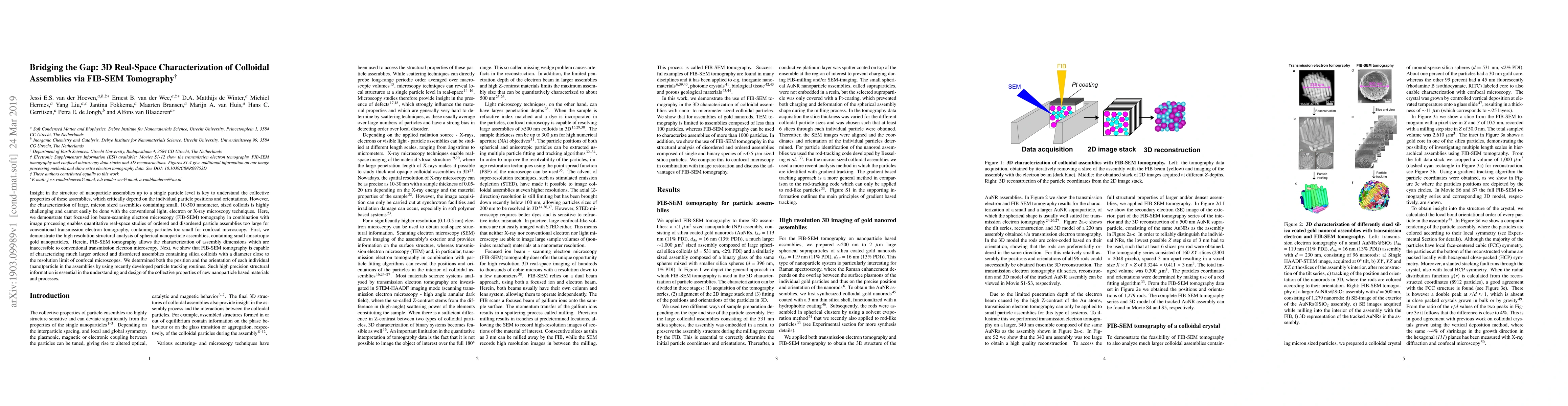
Insight in the structure of nanoparticle assemblies up to a single particle level is key to understand the collective properties of these assemblies, which critically depend on the individual particle positions and orientations. However, the characterization of large, micron sized assemblies containing small, 10-500 nanometer, sized colloids is highly challenging and cannot easily be done with the conventional light, electron or X-ray microscopy techniques. Here, we demonstrate that focused ion beam-scanning electron microscopy (FIB-SEM) tomography in combination with image processing enables quantitative real-space studies of ordered and disordered particle assemblies too large for conventional transmission electron tomography, containing particles too small for confocal microscopy. First, we demonstrate the high resolution structural analysis of spherical nanoparticle assemblies, containing small anisotropic gold nanoparticles. Herein, FIB-SEM tomography allows the characterization of assembly dimensions which are inaccessible to conventional transmission electron microscopy. Next, we show that FIB-SEM tomography is capable of characterizing much larger ordered and disordered assemblies containing silica colloids with a diameter close to the resolution limit of confocal microscopes. We determined both the position and the orientation of each individual (nano)particle in the assemblies by using recently developed particle tracking routines. Such high precision structural information is essential in the understanding and design of the collective properties of new nanoparticle based materials and processes.

Citation & Reference Network Visualization

This graph shows how this paper is connected to other research. The current paper is shown in dark gray, papers that cite it are in green, and papers it references are in blue. You can:

Current Paper
Papers citing this work (Citations)
Papers cited by this work (References)

• Hover over any node to see the paper title

• Click a node to view that paper's details

• Drag nodes to rearrange the graph

• Use the zoom controls (bottom right) to zoom in/out

Citations and References

Title Authors Year Actions
Improved sensitivity for subsurface imaging by contact resonance atomic force microscopy using Fano peaks
Yuyang Wang, Mingyu Duan, Yuan-Liu Chen
2024
View Paper
Advances in Focused Ion Beam Tomography for Three-Dimensional Characterization in Materials Science
Francesco Mura, F. Cognigni, Matteo Ferroni, V. Morandi, Marco Rossi
2023
View Paper
Three-dimensional visualization of dentine occlusion based on FIB-SEM tomography
Xinye Chen, Kaleigh M. Ryan, Deon Hines, Long Pan, Ke Du, Shiyou Xu
2023
View Paper
Small-Angle X-ray Scattering Analysis of Colloidal Crystals and Replica Materials Made from l-Arginine-Stabilized Silica Nanoparticles.
P. Mahale, Byeongdu Lee, H. Y. Cheng, M. Segad, T. Mallouk
2022
View Paper
Recent Notable Approaches to Study Self‐Assembly of Nanoparticles with X‐Ray Scattering and Electron Microscopy
F. Schulz, I. Lokteva, W. Parak, F. Lehmkühler
2021
View Paper
3D test sample for the calibration and quality control of stimulated emission depletion (STED) and confocal microscopes
Ernest B. van der Wee, J. Fokkema, Chris L Kennedy, Marc del Pozo, D. de Winter, P.N.A. Speets, H. Gerritsen, A. van Blaaderen
2021
View Paper
Unraveling Structural Details in Ga-Pd SCALMS Systems Using Correlative Nano-CT, 360° Electron Tomography and Analytical TEM
Janis Wirth, S. Englisch, D. Drobek, B. Apeleo Zubiri, Mingjian Wu, N. Taccardi, N. Raman, P. Wasserscheid, E. Spiecker
2021
View Paper
Recent Advances in Binary Colloidal Crystals for Photonics and Porous Material Fabrication.
Zhengting Zhang, Guiyun Yi, Peng Li, Xiuxiu Zhang, Zhuoyan Wan, Xiaodong Wang, Chuanxiang Zhang, Yulong Zhang
2021
View Paper
Structural Control over Bimetallic Core–Shell Nanorods for Surface-Enhanced Raman Spectroscopy
J. V. D. van der Hoeven, Tian-Song Deng, Wiebke Albrecht, L. Olthof, M. V. van Huis, P. D. de Jongh, A. van Blaaderen
2021
View Paper
Acoustic subsurface-atomic force microscopy: Three-dimensional imaging at the nanoscale
H. J. Sharahi, M. Janmaleki, L. Tetard, Seonghwan Kim, H. Sadeghian, G. Verbiest
2021
View Paper
Comparison of segmentation algorithms for FIB/SEM tomography of porous polymers: Importance of image contrast for machine learning segmentation
Martin Čalkovský, E. Müller, M. Meffert, Nadejda Firman, Frederik Mayer, M. Wegener, D. Gerthsen
2021
View Paper
3D test sample for the calibration and quality control of super-resolution and confocal microscopes
Ernest B. van der Wee, J. Fokkema, Chris L Kennedy, Marc del Pozo, D. A. Matthijs, D. Winter, P.N.A. Speets, H. Gerritsen, A. Blaaderen
2020
View Paper
Correlative Laboratory Nano‐CT and 360° Electron Tomography of Macropore Structures in Hierarchical Zeolites
B. Apeleo Zubiri, Janis Wirth, D. Drobek, S. Englisch, T. Przybilla, Tobias Weissenberger, W. Schwieger, E. Spiecker
2020
View Paper
Characterization of structures of particles
K. Manikas, G. G. Vogiatzis, P. Anderson, M. Hütter
2020
View Paper
Layering of bidisperse charged nanoparticles in sedimentation.
Xufeng Xu, Maarten Biesheuvel, H. Cölfen, E. Spruijt
2020
View Paper
Structural Color of Colloidal Clusters as a Tool to Investigate Structure and Dynamics
Junwei Wang, Umair Sultan, E. S. Goerlitzer, Chrameh Fru Mbah, M. Engel, N. Vogel
2019
View Paper
A Journey Through the Landscapes of Small Particles in Binary Colloidal Assemblies: Unveiling Structural Transitions from Isolated Particles to Clusters upon Variation in Composition
Valeria Lotito, T. Zambelli
2019
View Paper
Preparation and self-assembly of NaYF4:Eu3+ nanocrystals-PNIPAm complex nanogels
Dong-Ming Liang, Qiusheng Song, Ben Liu, Haihong Ma, Ziwen Zhang, Qiancheng Zhang
2020
View Paper

Similar Papers

Liquid phase fast electron tomography unravels the true 3D structure of colloidal assemblies

Da Wang, Sara Bals, Daniel Arenas Esteban, Ajinkya Kadu, Noa Olluyn, Ana Sánchez Iglesias, Alejandro Gomez Perez, Jesus Gonzalez Casablanca, Stavros Nicolopoulos, Luis M. Liz-Marzán

Published: 2023-11-27

Classification of FIB/SEM-tomography images for highly porous multiphase materials using random forest classifiers

Volker Schmidt, Matthias Neumann, Joachim R. Binder, Markus Osenberg, Ingo Manke, André Hilger, Nicole Bohn, Amalia Wagner, John Banhart

Published: 2022-07-29

Three-dimensional characterization of the steel-concrete interface by FIB-SEM nanotomography

Zhidong Zhang, Ueli M. Angst, O. Burkan Isgor, Nicolas Ruffray, Thilo Schmid

Published: 2023-10-09

The Potential of Subsampling and Inpainting for Fast Low-Dose Cryo FIB-SEM Imaging and Tomography

Damien McGrouther, Amirafshar Moshtaghpour, Daniel Nicholls, Alex W. Robinson, Angus I. Kirkland, Nigel D. Browning, Jack Wells, Maryna Kobylysnka, Zoe Broad, Roland A. Fleck

Published: 2023-10-30

Comments (0)

This website uses cookies to ensure you get the best experience. Learn more