This study evaluates various machine learning labeling strategies for detecting prostate cancer on MRI, finding that models trained with digital pathologist labels, which predict pixel-level Gleason patterns, achieve superior performance compared to those trained with radiologist or pathologist labels. These digital labels can enhance the reliability of machine learning models and reduce human annotation challenges.
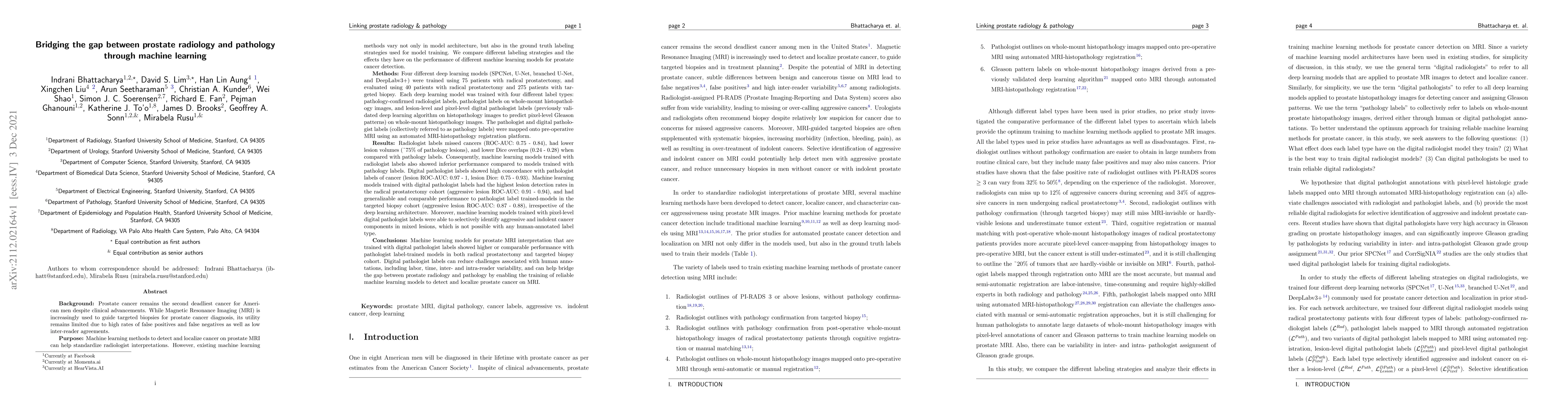
Prostate cancer is the second deadliest cancer for American men. While Magnetic Resonance Imaging (MRI) is increasingly used to guide targeted biopsies for prostate cancer diagnosis, its utility remains limited due to high rates of false positives and false negatives as well as low inter-reader agreements. Machine learning methods to detect and localize cancer on prostate MRI can help standardize radiologist interpretations. However, existing machine learning methods vary not only in model architecture, but also in the ground truth labeling strategies used for model training. In this study, we compare different labeling strategies, namely, pathology-confirmed radiologist labels, pathologist labels on whole-mount histopathology images, and lesion-level and pixel-level digital pathologist labels (previously validated deep learning algorithm on histopathology images to predict pixel-level Gleason patterns) on whole-mount histopathology images. We analyse the effects these labels have on the performance of the trained machine learning models. Our experiments show that (1) radiologist labels and models trained with them can miss cancers, or underestimate cancer extent, (2) digital pathologist labels and models trained with them have high concordance with pathologist labels, and (3) models trained with digital pathologist labels achieve the best performance in prostate cancer detection in two different cohorts with different disease distributions, irrespective of the model architecture used. Digital pathologist labels can reduce challenges associated with human annotations, including labor, time, inter- and intra-reader variability, and can help bridge the gap between prostate radiology and pathology by enabling the training of reliable machine learning models to detect and localize prostate cancer on MRI.
Get AI-generated insights about this paper's methodology, results, and significance.
Current paper (gray), citations (green), references (blue)
No similar papers found for this research.
Title | Authors | Year | Actions |
---|---|---|---|
The Paige Prostate Suite: Assistive Artificial Intelligence for Prostate Cancer Diagnosis
|
Cadth
|
2024
|
View Paper |
Framework for Deep Learning Based Multi-Modality Image Registration of Snapshot and Pathology Images
|
R. A. Schoop, L.M. de Roode, L. D. de Boer, B. Dashtbozorg
|
2024
|
View Paper |
Advantage of whole-mount histopathology in prostate cancer: current applications and future prospects
|
Lewei Duan, Zheng Liu, Fangning Wan, Bo Dai
|
2024
|
View Paper |
ArtHiFy: artificial histopathology-style features for improving MRI-based prostate cancer detection
|
Indrani Bhattacharya, Wei Shao, Xinran Li, S. Soerensen, Richard E. Fan, P. Ghanouni, James D. Brooks, G. Sonn, M. Rusu
|
2024
|
View Paper |
Boosting Few-Shot Learning with Disentangled Self-Supervised Learning and Meta-Learning for Medical Image Classification
|
Eva Pachetti, S. Tsaftaris, S. Colantonio
|
2024
|
View Paper |
A noninvasive method for predicting clinically significant prostate cancer using magnetic resonance imaging combined with PRKY promoter methylation level: a machine learning study
|
Yufei Wang, Weifeng Liu, Zeyu Chen, Yachen Zang, Lijun Xu, Zheng Dai, Yibin Zhou, Jin Zhu
|
2024
|
View Paper |
Identification and Localization of Indolent and Aggressive Prostate Cancers Using Multilevel Bi-LSTM.
|
Afnan M. Alhassan
|
2024
|
View Paper |
RAPHIA: A deep learning pipeline for the registration of MRI and whole-mount histopathology images of the prostate
|
Wei Shao, Sulaiman Vesal, S. Soerensen, Indrani Bhattacharya, Negar Golestani, Rikiya Yamashita, C. Kunder, Richard E. Fan, P. Ghanouni, James D. Brooks, G. Sonn, M. Rusu
|
2024
|
View Paper |
Deep learning model for the detection of prostate cancer and classification of clinically significant disease using multiparametric MRI in comparison to PI-RADs score.
|
Chunguang Yang, Basen Li, Yang Luan, Shiwei Wang, Yang Bian, Junbiao Zhang, Zefeng Wang, Bo Liu, Xin Chen, Marcus Hacker, Zhen Li, Xiang Li, Zhihua Wang
|
2024
|
View Paper |
Multi-scale statistical deformation based co-registration of prostate MRI and post-surgical whole mount histopathology.
|
Lin Li, R. Shiradkar, Noah Gottlieb, C. Buzzy, Amogh Hiremath, V. Viswanathan, G. MacLennan, Danly Omil Lima, K. Gupta, D. Shen, S. Tirumani, C. Magi-Galluzzi, A. Purysko, A. Madabhushi
|
2023
|
View Paper |
Application of Machine Learning and Deep Learning Models in Prostate Cancer Diagnosis Using Medical Images: A Systematic Review
|
O. Olabanjo, A. Wusu, Mauton Asokere, Oseni Taiwo Afisi, Basheerat Okugbesan, O. Olabanjo, O. Folorunso, Manuel Mazzara
|
2023
|
View Paper |
Comparison of MRI-Based Staging and Pathologic Staging for Predicting Biochemical Recurrence of Prostate Cancer After Radical Prostatectomy.
|
Katie M Merriman, S. Harmon, Mason J Belue, Enis Cagatay Yilmaz, Z. Blake, Nathan S. Lay, Tim E. Phelps, Maria J Merino, H. Parnes, Y. Law, S. Gurram, B. Wood, P. Choyke, P. Pinto, B. Turkbey
|
2023
|
View Paper |
Ultrasonographic pathological grading of prostate cancer using automatic region-based Gleason grading network
|
X. Lu, Shulian Zhang, Zhiyong Liu, Shaopeng Liu, Jun Huang, Guoquan Kong, Mingzhu Li, Yinying Liang, Yunneng Cui, Chuan Yang, Shen Zhao
|
2022
|
View Paper |
Correlated Feature Aggregation by Region Helps Distinguish Aggressive from Indolent Clear Cell Renal Cell Carcinoma Subtypes on CT
|
Karin Stacke, Indrani Bhattacharya, J. Tse, J. Brooks, G. Sonn, M. Rusu
|
2022
|
View Paper |
A review of artificial intelligence in prostate cancer detection on imaging
|
Indrani Bhattacharya, Y. Khandwala, Sulaiman Vesal, Wei Shao, Qianye Yang, S. Soerensen, Richard E. Fan, P. Ghanouni, C. Kunder, J. Brooks, Yipeng Hu, M. Rusu, G. Sonn
|
2022
|
View Paper |
Comments (0)