Bright 4B: Scaling Hyperspherical Learning for Segmentation in 3D Brightfield Microscopy
Publication
Metrics
Paper Preview
Abstract
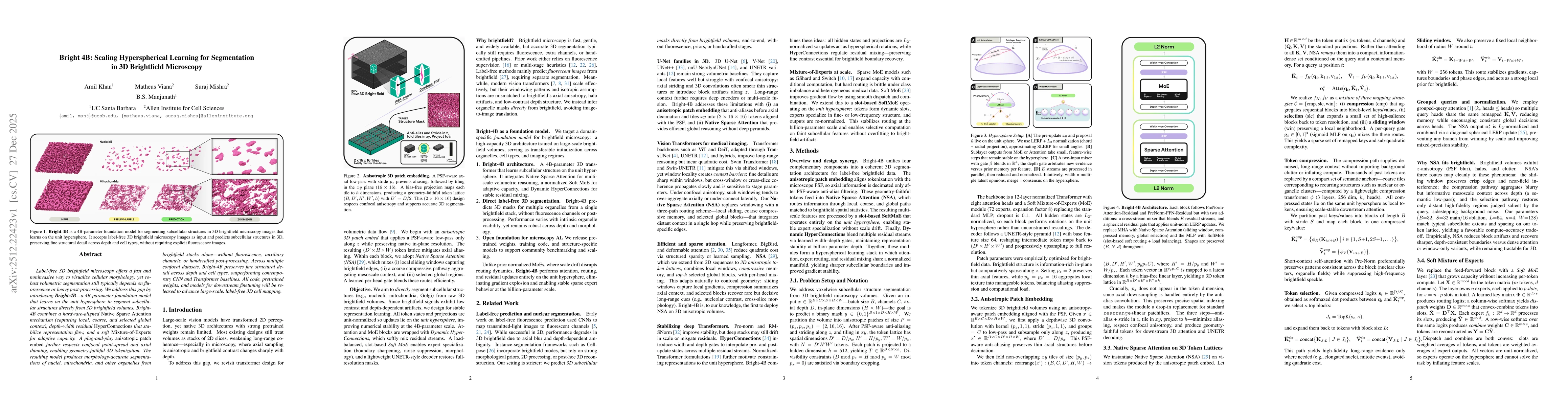
Label-free 3D brightfield microscopy offers a fast and noninvasive way to visualize cellular morphology, yet robust volumetric segmentation still typically depends on fluorescence or heavy post-processing. We address this gap by introducing Bright-4B, a 4 billion parameter foundation model that learns on the unit hypersphere to segment subcellular structures directly from 3D brightfield volumes. Bright-4B combines a hardware-aligned Native Sparse Attention mechanism (capturing local, coarse, and selected global context), depth-width residual HyperConnections that stabilize representation flow, and a soft Mixture-of-Experts for adaptive capacity. A plug-and-play anisotropic patch embed further respects confocal point-spread and axial thinning, enabling geometry-faithful 3D tokenization. The resulting model produces morphology-accurate segmentations of nuclei, mitochondria, and other organelles from brightfield stacks alone--without fluorescence, auxiliary channels, or handcrafted post-processing. Across multiple confocal datasets, Bright-4B preserves fine structural detail across depth and cell types, outperforming contemporary CNN and Transformer baselines. All code, pretrained weights, and models for downstream finetuning will be released to advance large-scale, label-free 3D cell mapping.
AI Key Findings
Get AI-generated insights about this paper's methodology, results, significance, and more — seven facets brought into focus.
Discussion 0