Publication
Metrics
AI Quick Summary
A new approach using ray tracing to analyze bright-field microscopy of transparent objects has been developed, offering two methods to extract quantitative phase information from images.
Paper Preview
Abstract
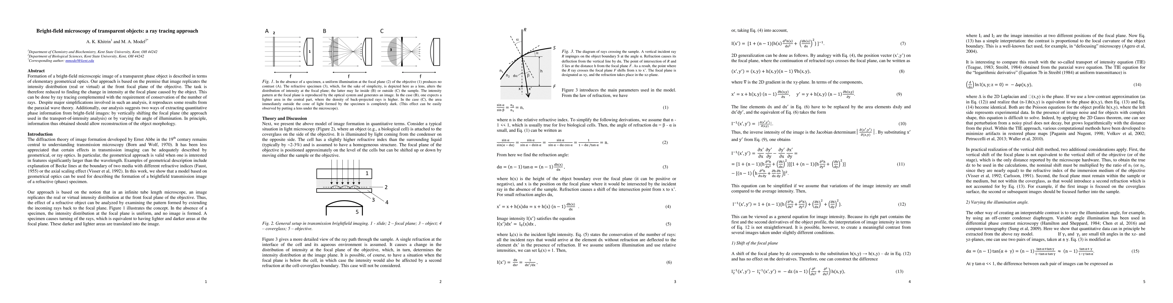
Formation of a bright-field microscopic image of a transparent phase object is described in terms of elementary geometrical optics. Our approach is based on the premise that image replicates the intensity distribution (real or virtual) at the front focal plane of the objective. The task is therefore reduced to finding the change in intensity at the focal plane caused by the object. This can be done by ray tracing complemented with the requirement of conservation of the number of rays. Despite major simplifications involved in such an analysis, it reproduces some results from the paraxial wave theory. Additionally, our analysis suggests two ways of extracting quantitative phase information from bright-field images: by vertically shifting the focal plane (the approach used in the transport-of-intensity analysis) or by varying the angle of illumination. In principle, information thus obtained should allow reconstruction of the object morphology.
AI Key Findings
Get AI-generated insights about this paper's methodology, results, significance, and more — seven facets brought into focus.
Impact
Paper Details
PDF Preview
Key Terms
Citation Network
Current paper (gray), citations (green), references (blue)
Display is limited for performance on very large graphs.
Discussion 0