Summary
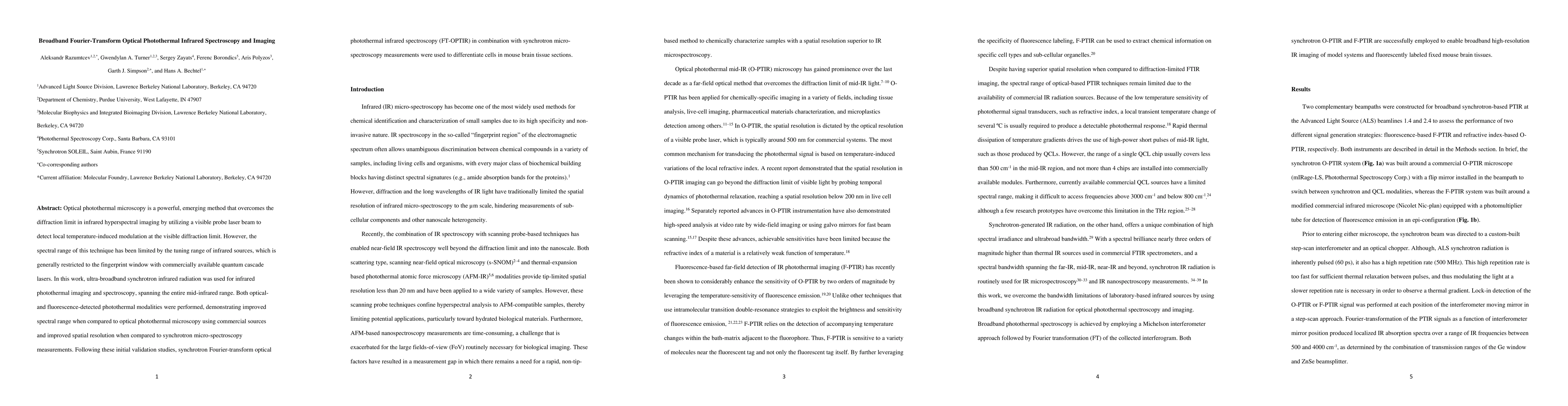
Optical photothermal microscopy is a powerful, emerging method that overcomes the diffraction limit in infrared hyperspectral imaging by utilizing a visible probe laser beam to detect local temperature-induced modulation at the visible diffraction limit. However, the spectral range of this technique has been limited by the tuning range of infrared sources, which is generally restricted to the fingerprint window with commercially available quantum cascade lasers. In this work, ultra-broadband synchrotron infrared radiation was used for infrared photothermal imaging and spectroscopy, spanning the entire mid-infrared range. Both optical- and fluorescence-detected photothermal modalities were performed, demonstrating improved spectral range when compared to optical photothermal microscopy using commercial sources and improved spatial resolution when compared to synchrotron micro-spectroscopy measurements. Following these initial validation studies, synchrotron Fourier-transform optical photothermal infrared spectroscopy (FT-OPTIR) in combination with synchrotron micro-spectroscopy measurements were used to differentiate cells in mouse brain tissue sections.
AI Key Findings
Get AI-generated insights about this paper's methodology, results, and significance.
Paper Details
PDF Preview
Key Terms
Citation Network
Current paper (gray), citations (green), references (blue)
Display is limited for performance on very large graphs.
Similar Papers
Found 4 papersNo citations found for this paper.
Comments (0)