The BT-Unet framework employs self-supervised learning with Barlow Twins to pre-train U-Net models for biomedical image segmentation, reducing the need for extensive annotated data. It achieves significant performance improvements even with limited labeled samples.
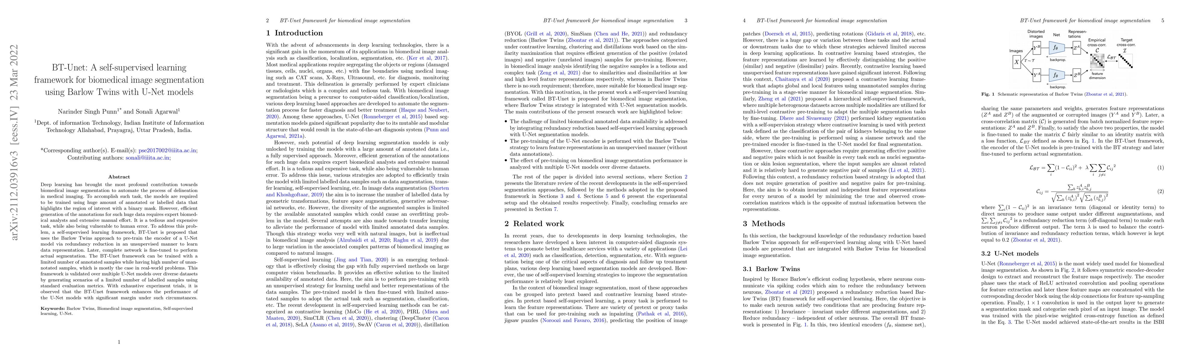
Deep learning has brought the most profound contribution towards biomedical image segmentation to automate the process of delineation in medical imaging. To accomplish such task, the models are required to be trained using huge amount of annotated or labelled data that highlights the region of interest with a binary mask. However, efficient generation of the annotations for such huge data requires expert biomedical analysts and extensive manual effort. It is a tedious and expensive task, while also being vulnerable to human error. To address this problem, a self-supervised learning framework, BT-Unet is proposed that uses the Barlow Twins approach to pre-train the encoder of a U-Net model via redundancy reduction in an unsupervised manner to learn data representation. Later, complete network is fine-tuned to perform actual segmentation. The BT-Unet framework can be trained with a limited number of annotated samples while having high number of unannotated samples, which is mostly the case in real-world problems. This framework is validated over multiple U-Net models over diverse datasets by generating scenarios of a limited number of labelled samples using standard evaluation metrics. With exhaustive experiment trials, it is observed that the BT-Unet framework enhances the performance of the U-Net models with significant margin under such circumstances.
This graph shows how this paper is connected to other research. The current paper is shown in dark gray, papers that cite it are in green, and papers it references are in blue. You can:
• Hover over any node to see the paper title
• Click a node to view that paper's details
• Drag nodes to rearrange the graph
• Use the zoom controls (bottom right) to zoom in/out
Title | Authors | Year | Actions |
---|---|---|---|
A survey on self-supervised methods for visual representation learning
|
Tobias Uelwer, Jan Robine, Stefan Sylvius Wagner, Marc Höftmann, Eric Upschulte, S. Konietzny, Maike Behrendt, Stefan Harmeling
|
2025
|
View Paper |
Maximising Histopathology Segmentation using Minimal Labels via Self-Supervision
|
Zeeshan Nisar, Thomas Lampert
|
2024
|
View Paper |
Small Intestine Tumor CT Image Segmentation Method Based on Self-Supervised Learning
|
Linpeng Li, Mingjia Wang
|
2024
|
View Paper |
A survey of vision-based condition monitoring methods using deep learning: A synthetic fiber rope perspective
|
A. Rani, D. O. Arroyo, P. Durdevic
|
2024
|
View Paper |
Devil is in Details: Locality-Aware 3D Abdominal CT Volume Generation for Self-Supervised Organ Segmentation
|
Yuran Wang, Zhijing Wan, Yansheng Qiu, Zheng Wang
|
2024
|
View Paper |
Dense Self-Supervised Learning for Medical Image Segmentation
|
Maxime Seince, L. L. Folgoc, Luiz Augusto Facury de Souza, Elsa Angelini
|
2024
|
View Paper |
Towards the Detection of Various and Extremely Small Wires in Cables
|
Daiki Takezaki, Chonho Lee, Yutaka Hoshina
|
2024
|
View Paper |
Light-Weight Semantic Segmentation Based on Vision Transformer and Self-Supervised Learning
|
Hsiu-Ming Lin, Guo-Shiang Lin, Chuan-Wang Chang, Ku-Yaw Chang, Wei-Cheng Yeh
|
2024
|
View Paper |
A Review of Semantic Medical Image Segmentation Based on Different Paradigms
|
Jianquan Tan, Wenrui Zhou, Ling Lin, Huxidan Jumahong
|
2024
|
View Paper |
2.5D Deep Learning Approach for Precise Image Segmentation of Gastro-Intestinal Tract
|
Jayanth Sai Marreddi, N. S. Punn
|
2024
|
View Paper |
A Review of various Architectures for Image Segmentation
|
Nagamani Gonthina, Narasimha Prasad
|
2024
|
View Paper |
STAIG: Spatial transcriptomics analysis via image-aided graph contrastive learning for domain exploration and alignment-free integration
|
Yitao Yang, Yang Cui, Xin Zeng, Yubo Zhang, Martin Loza, Sung-Joon Park, Kenta Nakai
|
2024
|
View Paper |
Knowledge-based U-Net and transfer learning for automatic boundary segmentation
|
Xiaoqi Zhou, Peixin Shi, B. Sheil, S. Suryasentana
|
2024
|
View Paper |
Thyroid ultrasound diagnosis improvement via multi-view self-supervised learning and two-stage pre-training
|
Jian Wang, Xin Yang, X. Jia, Wufeng Xue, Rusi Chen, Yanlin Chen, Xiliang Zhu, Lian Liu, Yan Cao, Jianqiao Zhou, Dong Ni, Ning Gu
|
2024
|
View Paper |
LWU-Net approach for Efficient Gastro-Intestinal Tract Image Segmentation in Resource-Constrained Environments
|
Marreddi Jayanth Sai, Singh Punn
|
2023
|
View Paper |
Auto-pore segmentation of digital microscopic leather images for species identification
|
Anjli Varghese, Sahil Jain, M. Jawahar, A. Prince
|
2023
|
View Paper |
Hybrid attention mechanism of feature fusion for medical image segmentation
|
Shanshan Tong, Zhentao Zuo, Zuxiang Liu, Dengdi Sun, Tiangang Zhou
|
2023
|
View Paper |
Deep Neural Networks for Brain Tumor Image Segmentation and Detection
|
Ritesh Chandra, Sadhana Tiwari, Shashi Shekhar Kumar, Sonali Agarwal, M. Syafrullah, K. Adiyarta
|
2023
|
View Paper |
Portable Skin Lesion Segmentation System with Accurate Lesion Localization Based on Weakly Supervised Learning
|
Hai Qin, Zhanjin Deng, Liye Shu, Yi Yin, Jintao Li, Li Zhou, Hui Zeng, Qiaokang Liang
|
2023
|
View Paper |
BTSeg: Barlow Twins Regularization for Domain Adaptation in Semantic Segmentation
|
Johannes Künzel, A. Hilsmann, P. Eisert
|
2023
|
View Paper |
A Comprehensive Overview of Computational Nuclei Segmentation Methods in Digital Pathology
|
Vasileios Magoulianitis, Catherine A. Alexander, C.-C. Jay Kuo
|
2023
|
View Paper |
Self-Supervised Deep-Learning Segmentation of Corneal Endothelium Specular Microscopy Images
|
S. Sánchez, Kevin D. Mendoza, Fernando J. Quintero, A. M. Prada, A. Tello, V. Galvis, L. Romero, A. Marrugo
|
2023
|
View Paper |
Deep neural networks for evaluation of specular microscopy images of the corneal endothelium with Fuchs' dystrophy
|
S. Sanchez, Kevin D. Mendoza, Fernando J. Quintero, A. M. Prada, A. Tello, V. Galvis, L. Romero, A. Marrugo
|
2023
|
View Paper |
DRS-UNET: A Deep Learning Approach for Diabetic Retinopathy Detection and Segmentation from Fundus Images
|
Ms. Renuka, Sandeep Gound, Dr. B. Meenakshi Sundaram, Santhosh Krishna, Peerzada Anzar Azmat, Malik Najeeb, Ul Habib, Avni Garg
|
2023
|
View Paper |
Brain Hemisphere Dissimilarity, a Self-Supervised Learning Approach for alpha-synucleinopathies prediction with FDG PET
|
Shikha Tripathi, P. Mattioli, C. Liguori, A. Chiaravalloti, D. Arnaldi, Luca Giancardo
|
2023
|
View Paper |
MV–MR: Multi-Views and Multi-Representations for Self-Supervised Learning and Knowledge Distillation
|
Vitaliy Kinakh, M. Drozdova, S. Voloshynovskiy
|
2023
|
View Paper |
A Review of Predictive and Contrastive Self-supervised Learning for Medical Images
|
Wei-Chien Wang, E. Ahn, Da-wei Feng, Jinman Kim
|
2023
|
View Paper |
Envisioning Reliable Diagnosis of Diabetic Retinopathy using U-Net Architecture
|
Shanmuga Priya, Dr. B. Meenakshi Sundaram, Peerzada Anzar Azmat, Avni Garg, Malik Najeeb, Ul Habib
|
2022
|
View Paper |
Towards the Identification and Classification of Solar Granulation Structures Using Semantic Segmentation
|
S. M. Díaz Castillo, A. Asensio Ramos, C. E. Fischer, S. Berdyugina
|
2022
|
View Paper |
Modality specific U-Net variants for biomedical image segmentation: a survey
|
N. S. Punn, Sonali Agarwal
|
2021
|
View Paper |
Published: 2024-09-17
Bjorn W. Schuller, Pancham Shukla, Harry Coppock, Jonah Anton
Published: 2022-12-26
Maxime Seince, Loic Le Folgoc, Luiz Augusto Facury de Souza, Elsa Angelini
Published: 2024-07-29
Comments (0)