Summary
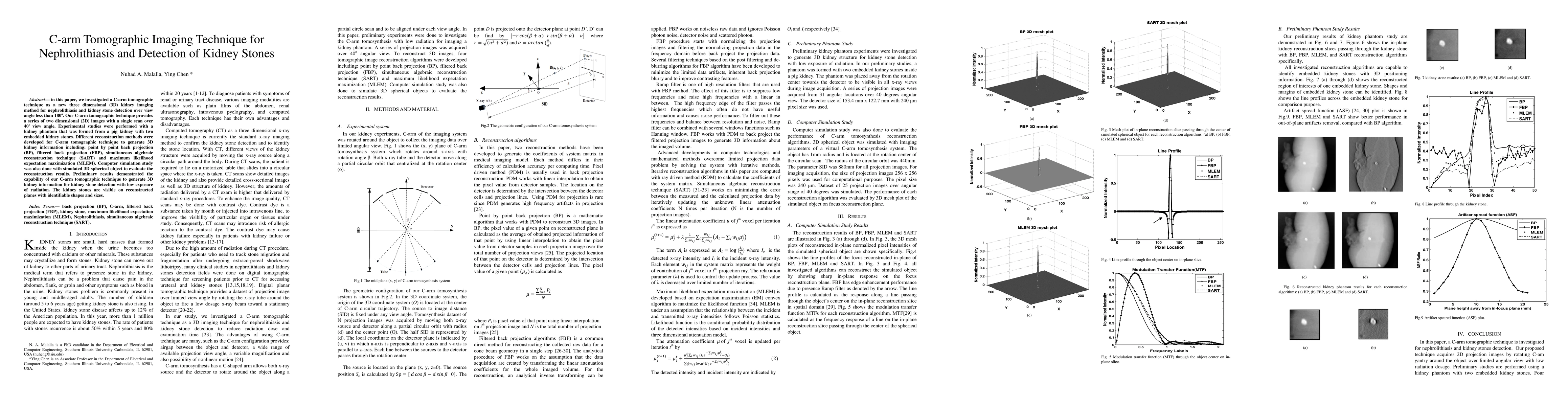
In this paper, we investigated a C-arm tomographic technique as a new three dimensional (3D) kidney imaging method for nephrolithiasis and kidney stone detection over view angle less than 180o. Our C-arm tomographic technique provides a series of two dimensional (2D) images with a single scan over 40o view angle. Experimental studies were performed with a kidney phantom that was formed from a pig kidney with two embedded kidney stones. Different reconstruction methods were developed for C-arm tomographic technique to generate 3D kidney information including: point by point back projection (BP), filtered back projection (FBP), simultaneous algebraic reconstruction technique (SART) and maximum likelihood expectation maximization (MLEM). Computer simulation study was also done with simulated 3D spherical object to evaluate the reconstruction results. Preliminary results demonstrated the capability of our C-arm tomographic technique to generate 3D kidney information for kidney stone detection with low exposure of radiation. The kidney stones are visible on reconstructed planes with identifiable shapes and sizes.
AI Key Findings
Get AI-generated insights about this paper's methodology, results, and significance.
Paper Details
PDF Preview
Key Terms
Citation Network
Current paper (gray), citations (green), references (blue)
Display is limited for performance on very large graphs.
Similar Papers
Found 4 papersAI-Driven Framework for Automated Detection of Kidney Stones in CT Images: Integration of Deep Learning Architectures and Transformers.
Alshenaifi, Reem, Alqahtani, Yahya, Ma, Shabnam et al.
No citations found for this paper.
Comments (0)