Calculating Point Spread Functions: Methods, Pitfalls and Solutions
Publication
Metrics
AI Quick Summary
This paper introduces Fourier-based techniques for calculating vector Point Spread Functions (PSF) in fluorescence microscopy, addressing sampling pitfalls and energy conservation issues. The proposed methods are shown to be accurate, computationally efficient, and adaptable to various imaging modalities.
Paper Preview
Abstract
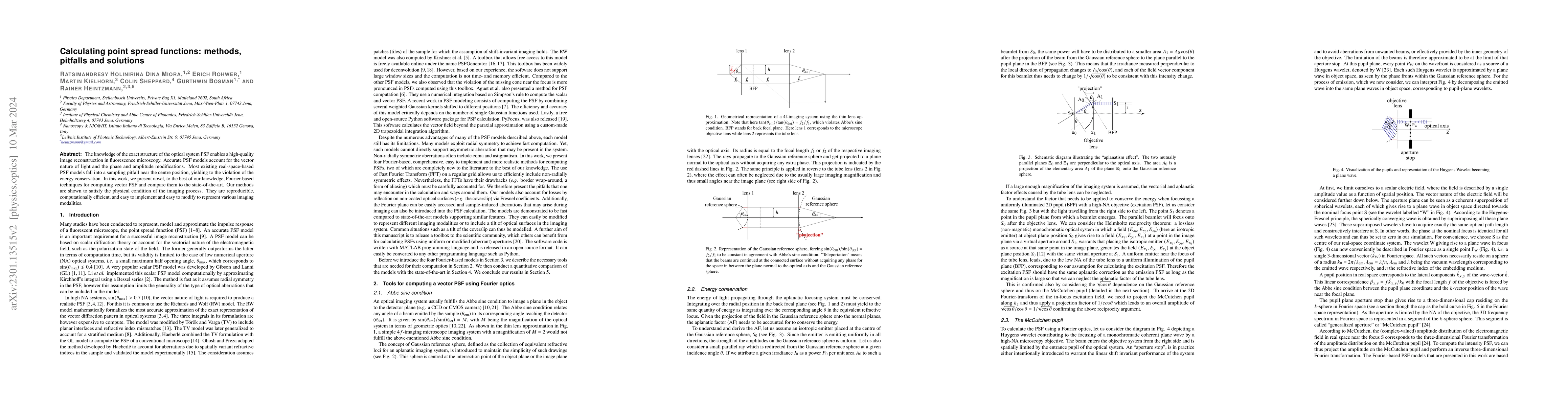
The knowledge of the exact structure of the optical system PSF enables a high-quality image reconstruction in fluorescence microscopy. Accurate PSF models account for the vector nature of light and the phase and amplitude modifications. Most existing real-space-based PSF models fall into a sampling pitfall near the centre position, yielding to the violation of the energy conservation. In this work, we present novel, to the best of our knowledge, Fourier-based techniques for computing vector PSF and compare them to the state-of-the-art. Our methods are shown to satisfy the physical condition of the imaging process. They are reproducible, computationally efficient, and easy to implement and easy to modify to represent various imaging modalities.
AI Key Findings
Get AI-generated insights about this paper's methodology, results, significance, and more — seven facets brought into focus.
Impact
Paper Details
Authors
PDF Preview
Key Terms
Citation Network
Current paper (gray), citations (green), references (blue)
Display is limited for performance on very large graphs.
Discussion 0