Publication
Metrics
AI Quick Summary
This paper introduces a cameraless high-throughput 3D imaging flow cytometry (3D-IFC) system that overcomes the projection problem of existing 2D imaging flow cytometry by using optical sectioning and light-sheet scanning to produce 3D cell images. The system achieves a throughput of 500 cells per second with sub-micron resolution in three dimensions, enabling more reliable cell phenotyping.
Paper Preview
Abstract
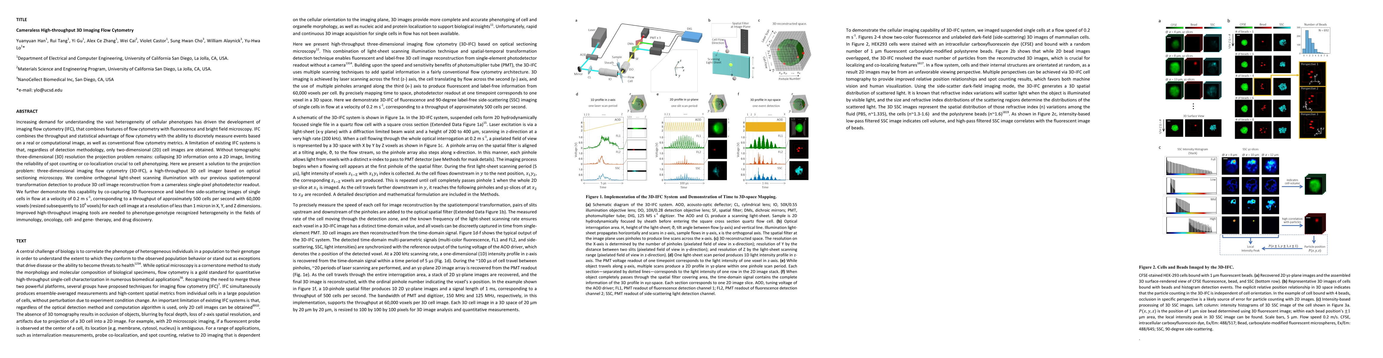
Increasing demand for understanding the vast heterogeneity of cellular phenotypes has driven the development of imaging flow cytometry (IFC), that combines features of flow cytometry with fluorescence and bright field microscopy. IFC combines the throughput and statistical advantage of flow cytometry with the ability to discretely measure events based on a real or computational image, as well as conventional flow cytometry metrics. A limitation of existing IFC systems is that, regardless of detection methodology, only two-dimensional (2D) cell images are obtained. Without tomographic three-dimensional (3D) resolution the projection problem remains: collapsing 3D information onto a 2D image, limiting the reliability of spot counting or co-localization crucial to cell phenotyping. Here we present a solution to the projection problem: three-dimensional imaging flow cytometry (3D-IFC), a high-throughput 3D cell imager based on optical sectioning microscopy. We combine orthogonal light-sheet scanning illumination with our previous spatiotemporal transformation detection to produce 3D cell image reconstruction from a cameraless single-pixel photodetector readout. We further demonstrate this capability by co-capturing 3D fluorescence and label-free side-scattering images of single cells in flow at a velocity of 0.2 m s-1, corresponding to a throughput of approximately 500 cells per second with 60,000 voxels (resized subsequently to 106 voxels) for each cell image at a resolution of less than 1 micron in X, Y, and Z dimensions. Improved high-throughput imaging tools are needed to phenotype-genotype recognized heterogeneity in the fields of immunology, oncology, cell- and gene- therapy, and drug discovery.
AI Key Findings
Get AI-generated insights about this paper's methodology, results, significance, and more — seven facets brought into focus.
Impact
Paper Details
PDF Preview
Key Terms
Citation Network
Current paper (gray), citations (green), references (blue)
Display is limited for performance on very large graphs.
Discussion 0