Publication
Metrics
AI Quick Summary
This paper demonstrates a cavity-enhanced Raman microscopy technique for studying individual carbon nanotubes, achieving a 320-times Raman signal enhancement and identifying structural parameters, promising for molecular diagnostics and nanomechanical control. The method also exhibits potential for higher enhancement and quantitative signal acquisition without intrinsic background noise.
Paper Preview
Abstract
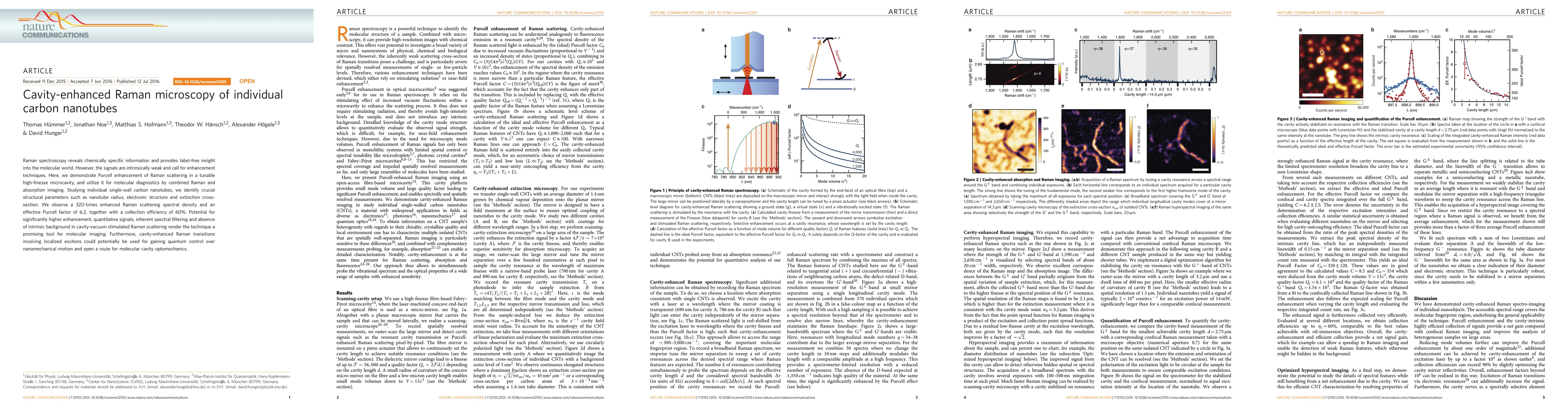
Raman spectroscopy reveals chemically specific information and provides label-free insight into the molecular world. However, the signals are intrinsically weak and call for enhancement techniques. Here, we demonstrate Purcell enhancement of Raman scattering in a tunable high-finesse microcavity, and utilize it for molecular diagnostics by combined Raman and absorption imaging. Studying individual single-wall carbon nanotubes, we identify crucial structural parameters such as nanotube radius, electronic structure and extinction cross-section. We observe a 320-times enhanced Raman scattering spectral density and an effective Purcell factor of 6.2, together with a collection efficiency of 60%. Potential for significantly higher enhancement, quantitative signals, inherent spectral filtering and absence of intrinsic background in cavity-vacuum stimulated Raman scattering render the technique a promising tool for molecular imaging. Furthermore, cavity-enhanced Raman transitions involving localized excitons could potentially be used for gaining quantum control over nanomechanical motion and open a route for molecular cavity optomechanics.
AI Key Findings
Get AI-generated insights about this paper's methodology, results, significance, and more — seven facets brought into focus.
Impact
Paper Details
PDF Preview
Key Terms
Citation Network
Current paper (gray), citations (green), references (blue)
Display is limited for performance on very large graphs.
Discussion 0