CE-Net: Context Encoder Network for 2D Medical Image Segmentation

Source: ArXiv

AI Quick Summary

CE-Net, a context encoder network, is proposed to improve 2D medical image segmentation by preserving spatial information and capturing high-level context. It outperforms U-Net and other methods in tasks like optic disc, vessel, lung, cell, and retinal layer segmentation.

Share: Share on X LinkedIn

Summary

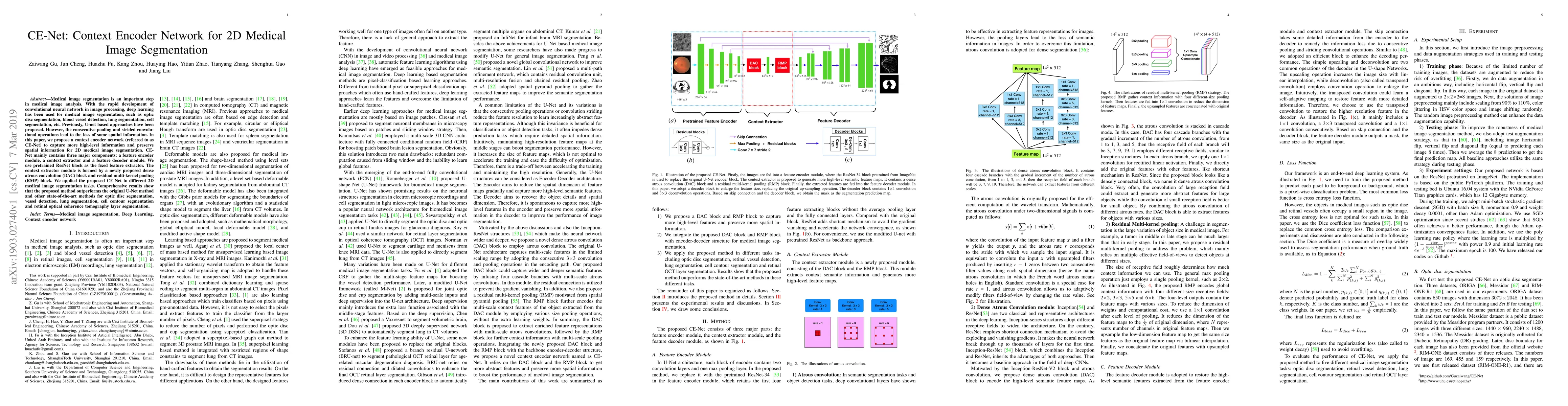
Medical image segmentation is an important step in medical image analysis. With the rapid development of convolutional neural network in image processing, deep learning has been used for medical image segmentation, such as optic disc segmentation, blood vessel detection, lung segmentation, cell segmentation, etc. Previously, U-net based approaches have been proposed. However, the consecutive pooling and strided convolutional operations lead to the loss of some spatial information. In this paper, we propose a context encoder network (referred to as CE-Net) to capture more high-level information and preserve spatial information for 2D medical image segmentation. CE-Net mainly contains three major components: a feature encoder module, a context extractor and a feature decoder module. We use pretrained ResNet block as the fixed feature extractor. The context extractor module is formed by a newly proposed dense atrous convolution (DAC) block and residual multi-kernel pooling (RMP) block. We applied the proposed CE-Net to different 2D medical image segmentation tasks. Comprehensive results show that the proposed method outperforms the original U-Net method and other state-of-the-art methods for optic disc segmentation, vessel detection, lung segmentation, cell contour segmentation and retinal optical coherence tomography layer segmentation.

AI Key Findings

Generated Sep 03, 2025

Methodology

The paper proposes CE-Net, a Context Encoder Network for 2D medical image segmentation, which incorporates a pretrained ResNet block, a dense atrous convolution (DAC) block, and a residual multi-kernel pooling (RMP) block to capture high-level semantic features while preserving spatial information.

Key Results

  • CE-Net outperforms U-Net and other state-of-the-art methods in optic disc segmentation, vessel detection, lung segmentation, cell contour segmentation, and retinal OCT layer segmentation.
  • The proposed method reduces overlapping error by 13.6% for lung segmentation and increases sensitivity by 6.8% for retinal vessel detection compared to the backbone model.
  • CE-Net achieves higher Dice coefficient scores than U-Net in cell contour segmentation.

Significance

This research presents a novel deep learning framework for medical image segmentation that preserves more spatial information and captures high-level semantic features, which can significantly improve the accuracy of various medical image segmentation tasks.

Technical Contribution

The introduction of dense atrous convolution (DAC) and residual multi-kernel pooling (RMP) blocks, integrated into a ResNet-modified U-Net structure, to effectively capture high-level semantic features and preserve spatial information for medical image segmentation.

Novelty

CE-Net differentiates itself from existing methods by addressing the loss of spatial information caused by consecutive pooling and strided convolutional operations in traditional U-Net architectures, thereby improving segmentation performance across multiple medical image segmentation tasks.

Limitations

  • The paper does not discuss the computational resources required for training the model.
  • No information is provided on the generalization of the model to 3D medical images.

Future Work

  • Explore the application of CE-Net to 3D medical image segmentation tasks.
  • Investigate the computational efficiency and scalability of the proposed method for real-time or large-scale medical image analysis.

Paper Details

PDF Preview

Download PDF

Key Terms

segmentation (0.307)
medical image (0.304)
net (0.245)
medical (0.239)
image (0.193)
block (0.193)
module (0.185)
disc (0.170)

Citation Network

Current paper (gray), citations (green), references (blue)

Display is limited for performance on very large graphs.

Current Paper
Citations
References
Click to view
Title Authors Year Actions

Comments (0)

This website uses cookies to ensure you get the best experience. Learn more