Cell divisions imprint long lasting elastic strain fields in epithelial tissues
Publication
Metrics
AI Quick Summary
This study analyzes the deformation fields induced by cell divisions in fruit fly wing epithelia, finding that the resulting elastic strain fields persist for up to 3.5 hours, corresponding to the tissue's fluidization timescale. The method reveals transient isotropic and traceless-symmetric force dipole fields from cell divisions, providing insights into tissue mechanics.
Paper Preview
Abstract
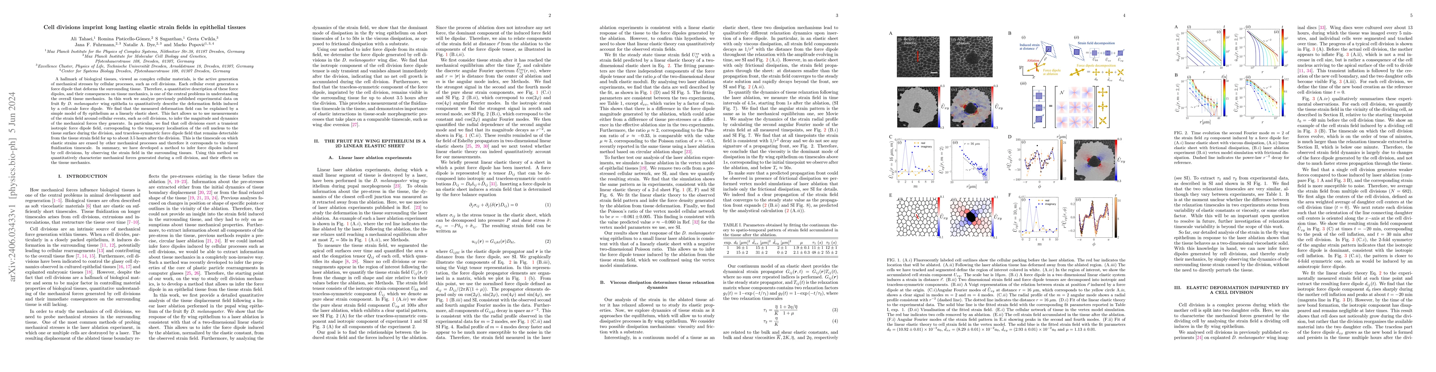
A hallmark of biological tissues, viewed as complex cellular materials, is the active generation of mechanical stresses by cellular processes, such as cell divisions. Each cellular event generates a force dipole that deforms the surrounding tissue. Therefore, a quantitative description of these force dipoles, and their consequences on tissue mechanics, is one of the central problems in understanding the overall tissue mechanics. In this work we analyze previously published experimental data on fruit fly \textit{D. melanogaster} wing epithelia to quantitatively describe the deformation fields induced by a cell-scale force dipole. We find that the measured deformation field can be explained by a simple model of fly epithelium as a linearly elastic sheet. This fact allows us to use measurements of the strain field around cellular events, such as cell divisions, to infer the magnitude and dynamics of the mechanical forces they generate. In particular, we find that cell divisions exert a transient isotropic force dipole field, corresponding to the temporary localisation of the cell nucleus to the tissue surface during the division, and traceless-symmetric force dipole field that remains detectable from the tissue strain field for up to about $3.5$ hours after the division. This is the timescale on which elastic strains are erased by other mechanical processes and therefore it corresponds to the tissue fluidization timescale. In summary, we have developed a method to infer force dipoles induced by cell divisions, by observing the strain field in the surrounding tissues. Using this method we quantitatively characterize mechanical forces generated during a cell division, and their effects on the tissue mechanics.
AI Key Findings
Get AI-generated insights about this paper's methodology, results, significance, and more — seven facets brought into focus.
Impact
Paper Details
Authors
PDF Preview
Key Terms
Citation Network
Current paper (gray), citations (green), references (blue)
Display is limited for performance on very large graphs.
Discussion 0