Cellular Micromasonry: Biofabrication with Single Cell Precision

Source: ArXiv

AI Quick Summary

This paper introduces a novel method for 3D cell assembly with single-cell precision, enabling the creation of complex tissue structures with detailed spatial heterogeneities. The technique demonstrates functional viability and opens new avenues for studying cellular behavior and interactions in defined contexts.

Share: Share on X LinkedIn

Summary

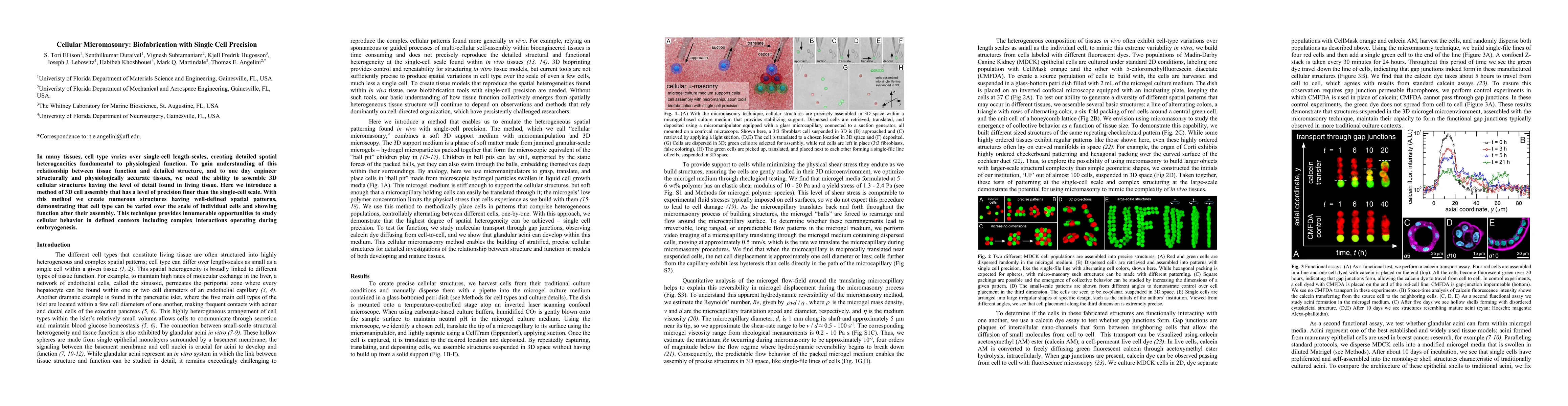
In many tissues, cell type varies over single-cell length-scales, creating detailed spatial heterogeneities fundamental to physiological function. To gain understanding of this relationship between tissue function and detailed structure, and to one day engineer structurally and physiologically accurate tissues, we need the ability to assemble 3D cellular structures having the level of detail found in living tissue. Here we introduce a method of 3D cell assembly that has a level of precision finer than the single-cell scale. With this method we create numerous structures having well-defined spatial patterns, demonstrating that cell type can be varied over the scale of individual cells and showing function after their assembly. This technique provides innumerable opportunities to study cellular behavior in defined contexts including complex interactions operating during embryogenesis.

AI Key Findings

Generated Sep 03, 2025

Methodology

The research introduces a method called 'micromasonry' for 3D cell assembly with single-cell precision, utilizing jammed microgel medium to create well-defined spatial patterns of cell types.

Key Results

  • Cellular structures with single-cell precision were successfully assembled.
  • Functional gap junctions were maintained in structures assembled with micromasonry.
  • MDCK cells self-assembled into monolayer shell structures resembling traditionally cultured acini.
  • Three-color, 3D structures including a layered pyramid and stack of 2x2 structures were built from MDCK cells.
  • A hollow spherical structure was assembled to approximate a glandular acinus.

Significance

This research provides a novel technique for studying cellular behavior in defined contexts, including complex interactions during embryogenesis, and opens up opportunities for engineering structurally and physiologically accurate tissues.

Technical Contribution

The introduction of a micromasonry technique for 3D cell assembly with single-cell precision, utilizing jammed microgel medium to create intricate cellular structures.

Novelty

This method offers a finer level of precision than previous techniques, enabling the creation of cellular structures with detailed spatial heterogeneities akin to living tissues.

Limitations

  • Current manual procedure limits the physical scale of structures that can be assembled.
  • The study did not explore long-term functionalities of the assembled structures.

Future Work

  • Automate the micromasonry process using engineering tools like 3D image segmentation, 3D cell tracking, and pick-and-place robotics.
  • Develop computational tools based on machine learning and AI for controlling machine operations in uncertain environments to build larger and more complex structures.

Paper Details

PDF Preview

Download PDF

Key Terms

cell (0.325)
cell (0.310)
assembly (0.261)
tissue (0.255)
cellular (0.250)
assembly (0.244)
cellular (0.241)
having (0.204)
Title Authors Year Actions

Comments (0)

This website uses cookies to ensure you get the best experience. Learn more