Cerebral oxygen extraction fraction MRI: techniques and applications
Publication
Metrics
AI Quick Summary
MRI-based methods have been developed to measure oxygen extraction fraction (OEF) in the human brain, offering non-invasive and radiation-free alternatives to 15O positron emission tomography.
Paper Preview
Abstract
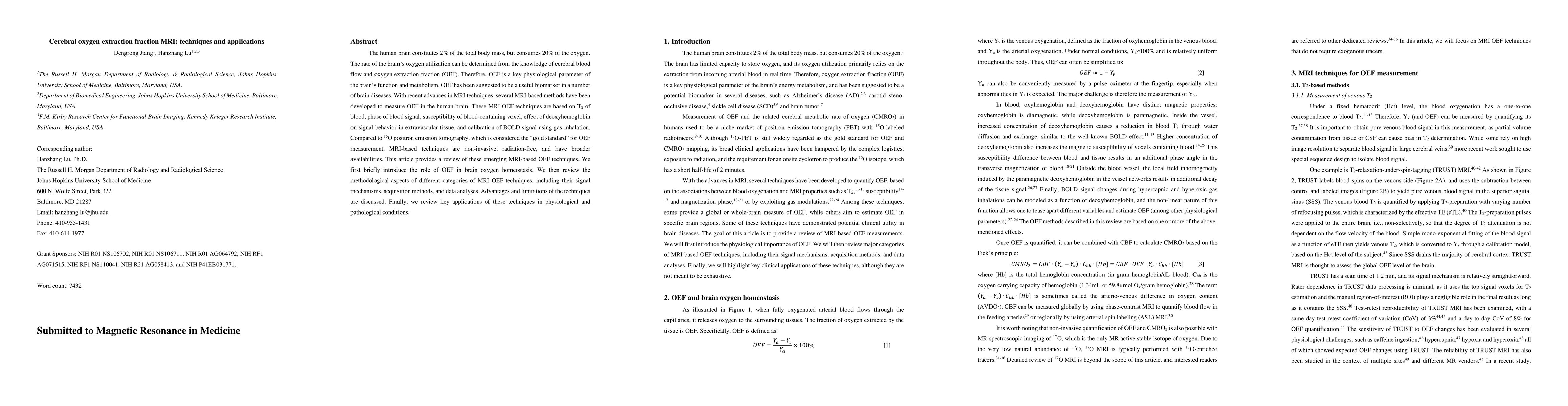
The human brain constitutes 2% of the total body mass, but consumes 20% of the oxygen. The rate of the brain's oxygen utilization can be determined from the knowledge of cerebral blood flow and oxygen extraction fraction (OEF). Therefore, OEF is a key physiological parameter of the brain's function and metabolism. OEF has been suggested to be a useful biomarker in a number of brain diseases. With recent advances in MRI techniques, several MRI-based methods have been developed to measure OEF in the human brain. These MRI OEF techniques are based on T2 of blood, phase of blood signal, susceptibility of blood-containing voxel, effect of deoxyhemoglobin on signal behavior in extravascular tissue, and calibration of BOLD signal using gas-inhalation. Compared to 15O positron emission tomography, which is considered the "gold standard" for OEF measurement, MRI-based techniques are non-invasive, radiation-free, and have broader availabilities. This article provides a review of these emerging MRI-based OEF techniques. We first briefly introduce the role of OEF in brain oxygen homeostasis. We then review the methodological aspects of different categories of MRI OEF techniques, including their signal mechanisms, acquisition methods, and data analyses. Advantages and limitations of the techniques are discussed. Finally, we review key applications of these techniques in physiological and pathological conditions.
AI Key Findings
Get AI-generated insights about this paper's methodology, results, significance, and more — seven facets brought into focus.
Impact
Paper Details
Authors
PDF Preview
Key Terms
Citation Network
Current paper (gray), citations (green), references (blue)
Display is limited for performance on very large graphs.
Discussion 0