CEST MR fingerprinting (CEST-MRF) for Brain Tumor Quantification Using EPI Readout and Deep Learning Reconstruction
Publication
Metrics
AI Quick Summary
This study developed a CEST-MRF method for brain tumor quantification using EPI readout and deep learning reconstruction, demonstrating its accuracy, reproducibility, and clinical utility in distinguishing tumor regions from healthy tissue.
Paper Preview
Abstract
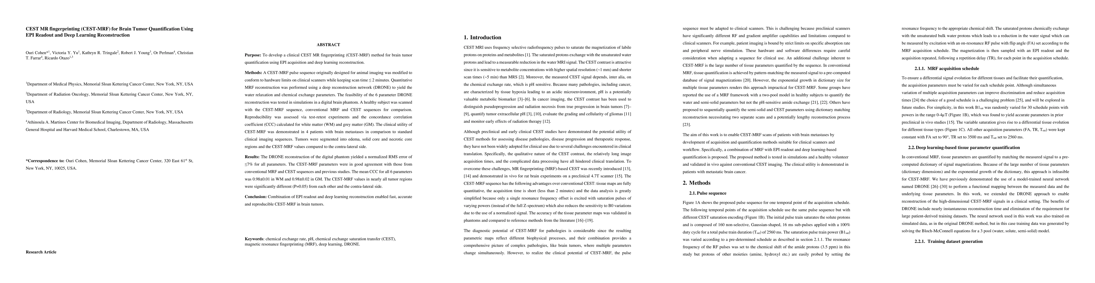
$\textbf{Purpose}$: To develop a clinical CEST MR fingerprinting (CEST-MRF) method for brain tumor quantification using EPI acquisition and deep learning reconstruction. $\textbf{Methods}$: A CEST-MRF pulse sequence originally designed for animal imaging was modified to conform to hardware limits on clinical scanners while keeping scan time $\leq$ 2 minutes. Quantitative MRF reconstruction was performed using a deep reconstruction network (DRONE) to yield the water relaxation and chemical exchange parameters. The feasibility of the 6 parameter DRONE reconstruction was tested in simulations in a digital brain phantom. A healthy subject was scanned with the CEST-MRF sequence, conventional MRF and CEST sequences for comparison. Reproducibility was assessed via test-retest experiments and the concordance correlation coefficient (CCC) calculated for white matter (WM) and grey matter (GM). The clinical utility of CEST-MRF was demonstrated in 4 patients with brain metastases in comparison to standard clinical imaging sequences. Tumors were segmented into edema, solid core and necrotic core regions and the CEST-MRF values compared to the contra-lateral side. $\textbf{Results}$: The DRONE reconstruction of the digital phantom yielded a normalized RMS error of $\leq$ 7% for all parameters. The CEST-MRF parameters were in good agreement with those from conventional MRF and CEST sequences and previous studies. The mean CCC for all 6 parameters was 0.98$\pm$0.01 in WM and 0.98$\pm$0.02 in GM. The CEST-MRF values in nearly all tumor regions were significantly different (P=0.05) from each other and the contra-lateral side. $\textbf{Conclusion}$: Combination of EPI readout and deep learning reconstruction enabled fast, accurate and reproducible CEST-MRF in brain tumors.
AI Key Findings
Get AI-generated insights about this paper's methodology, results, significance, and more — seven facets brought into focus.
Impact
Paper Details
Authors
PDF Preview
Key Terms
Citation Network
Current paper (gray), citations (green), references (blue)
Display is limited for performance on very large graphs.
Discussion 0