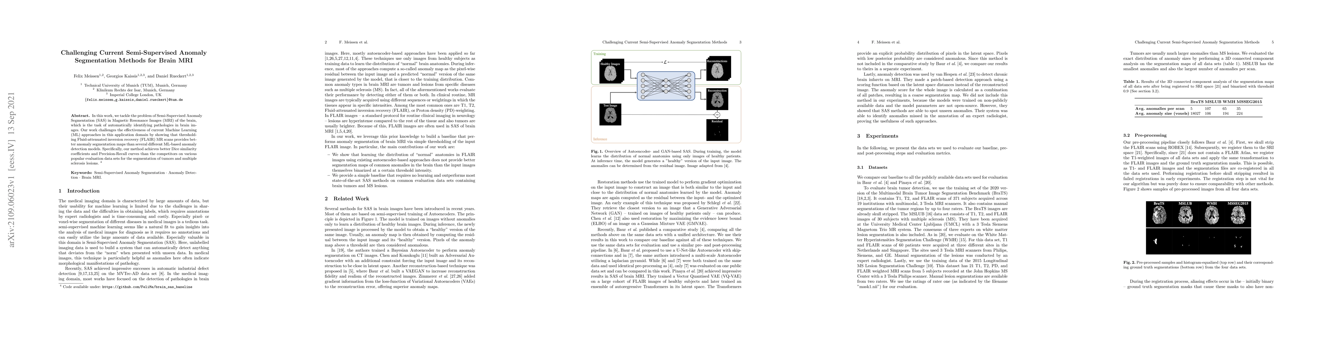
Publication
Metrics
AI Quick Summary
This paper challenges the effectiveness of current semi-supervised anomaly segmentation methods for brain MRI, demonstrating that thresholding FLAIR MR scans outperforms several machine learning models in identifying brain pathologies, achieving superior Dice similarity coefficients and Precision-Recall curves on standard datasets.
Paper Preview
Abstract
In this work, we tackle the problem of Semi-Supervised Anomaly Segmentation (SAS) in Magnetic Resonance Images (MRI) of the brain, which is the task of automatically identifying pathologies in brain images. Our work challenges the effectiveness of current Machine Learning (ML) approaches in this application domain by showing that thresholding Fluid-attenuated inversion recovery (FLAIR) MR scans provides better anomaly segmentation maps than several different ML-based anomaly detection models. Specifically, our method achieves better Dice similarity coefficients and Precision-Recall curves than the competitors on various popular evaluation data sets for the segmentation of tumors and multiple sclerosis lesions.
AI Key Findings
Get AI-generated insights about this paper's methodology, results, significance, and more — seven facets brought into focus.
Impact
Paper Details
Authors
PDF Preview
Key Terms
Citation Network
Current paper (gray), citations (green), references (blue)
Display is limited for performance on very large graphs.
Discussion 0