01
MethodologyHow they did it
A hybrid CNN-transformer architecture was proposed for medical image segmentation.
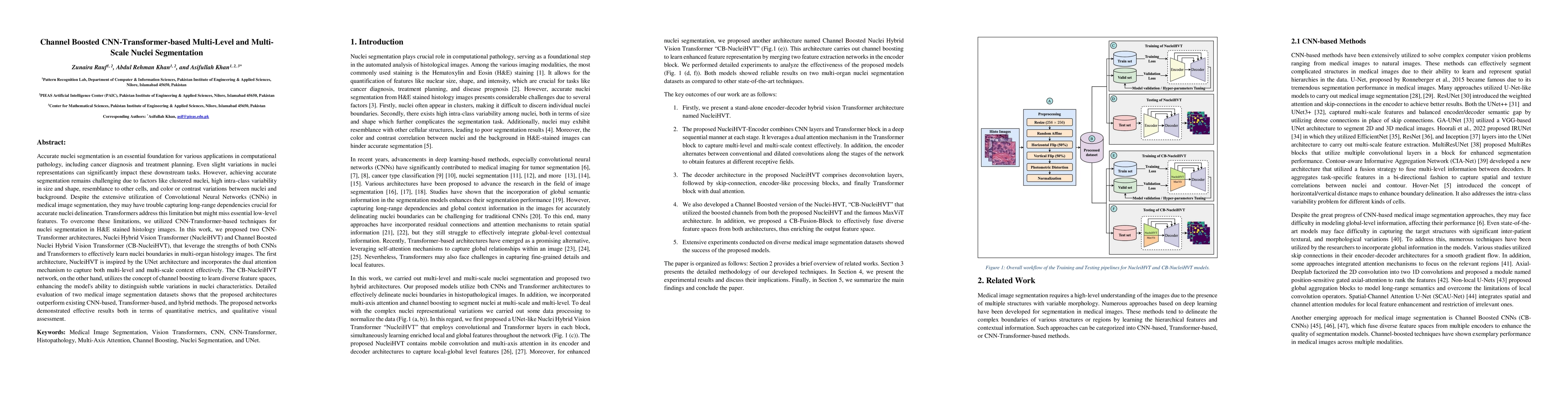
This research proposes two CNN-Transformer architectures, NucleiHVT and CB-NucleiHVT, to improve nuclei segmentation in H&E stained histology images by leveraging both CNNs and Transformers. The CB-NucleiHVT incorporates channel boosting to enhance feature differentiation, achieving superior performance over existing methods in multi-organ datasets.
This research proposes two CNN-Transformer architectures, NucleiHVT and CB-NucleiHVT, to improve nuclei segmentation in H&E stained histology images by leveraging both CNNs and Transformers. The CB-NucleiHVT incorporates channel boosting to enhance feature differentiation, achieving superior performance over existing methods in multi-organ datasets.
A hybrid CNN-transformer architecture was proposed for medical image segmentation. More in Methodology →
Improved accuracy compared to state-of-the-art methods — Enhanced robustness against various types of noise and artifacts More in Key Results →
The proposed method has the potential to improve diagnosis accuracy and reduce healthcare costs. More in Significance →
Requires large amounts of labeled data for training — May not generalize well to new, unseen medical images More in Limitations →
Accurate nuclei segmentation is an essential foundation for various applications in computational pathology, including cancer diagnosis and treatment planning. Even slight variations in nuclei representations can significantly impact these downstream tasks. However, achieving accurate segmentation remains challenging due to factors like clustered nuclei, high intra-class variability in size and shape, resemblance to other cells, and color or contrast variations between nuclei and background. Despite the extensive utilization of Convolutional Neural Networks (CNNs) in medical image segmentation, they may have trouble capturing long-range dependencies crucial for accurate nuclei delineation. Transformers address this limitation but might miss essential low-level features. To overcome these limitations, we utilized CNN-Transformer-based techniques for nuclei segmentation in H&E stained histology images. In this work, we proposed two CNN-Transformer architectures, Nuclei Hybrid Vision Transformer (NucleiHVT) and Channel Boosted Nuclei Hybrid Vision Transformer (CB-NucleiHVT), that leverage the strengths of both CNNs and Transformers to effectively learn nuclei boundaries in multi-organ histology images. The first architecture, NucleiHVT is inspired by the UNet architecture and incorporates the dual attention mechanism to capture both multi-level and multi-scale context effectively. The CB-NucleiHVT network, on the other hand, utilizes the concept of channel boosting to learn diverse feature spaces, enhancing the model's ability to distinguish subtle variations in nuclei characteristics. Detailed evaluation of two medical image segmentation datasets shows that the proposed architectures outperform existing CNN-based, Transformer-based, and hybrid methods. The proposed networks demonstrated effective results both in terms of quantitative metrics, and qualitative visual assessment.
Seven facets of this paper, analysed and brought into focus by AI.
The proposed method has the potential to improve diagnosis accuracy and reduce healthcare costs.
A hybrid CNN-transformer architecture was proposed for medical image segmentation.
The proposed method has the potential to improve diagnosis accuracy and reduce healthcare costs.
A novel attention mechanism was introduced to improve the performance of CNN-based models in medical image segmentation.
The proposed method combines the strengths of both CNNs and transformers, leading to improved accuracy and robustness.
Current paper (gray), citations (green), references (blue)
Display is limited for performance on very large graphs.
Discussion 0