Chest X-Ray Analysis of Tuberculosis by Deep Learning with Segmentation and Augmentation
Publication
Metrics
AI Quick Summary
This research explores the use of deep learning techniques, specifically convolutional neural networks (CNNs), for analyzing chest X-rays to detect tuberculosis. The study highlights the effectiveness of lung segmentation and data augmentation methods in improving the accuracy of computer-aided diagnosis (CADx) even with a small, unbalanced dataset. The findings suggest that lossless data augmentation yields the best results in terms of validation loss and accuracy.
Paper Preview
Abstract
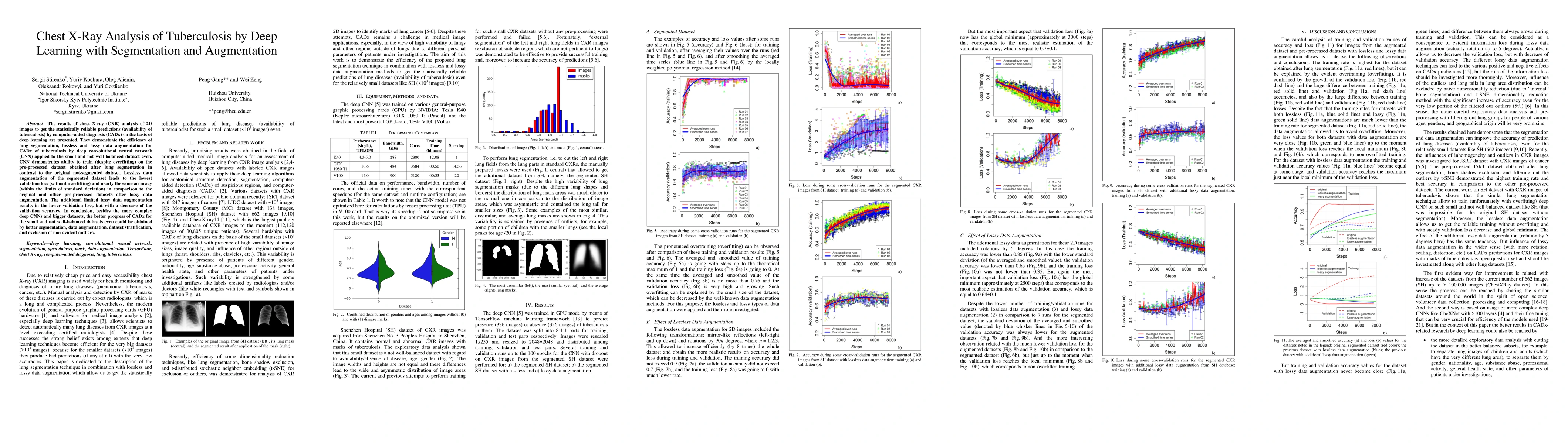
The results of chest X-ray (CXR) analysis of 2D images to get the statistically reliable predictions (availability of tuberculosis) by computer-aided diagnosis (CADx) on the basis of deep learning are presented. They demonstrate the efficiency of lung segmentation, lossless and lossy data augmentation for CADx of tuberculosis by deep convolutional neural network (CNN) applied to the small and not well-balanced dataset even. CNN demonstrates ability to train (despite overfitting) on the pre-processed dataset obtained after lung segmentation in contrast to the original not-segmented dataset. Lossless data augmentation of the segmented dataset leads to the lowest validation loss (without overfitting) and nearly the same accuracy (within the limits of standard deviation) in comparison to the original and other pre-processed datasets after lossy data augmentation. The additional limited lossy data augmentation results in the lower validation loss, but with a decrease of the validation accuracy. In conclusion, besides the more complex deep CNNs and bigger datasets, the better progress of CADx for the small and not well-balanced datasets even could be obtained by better segmentation, data augmentation, dataset stratification, and exclusion of non-evident outliers.
AI Key Findings
Get AI-generated insights about this paper's methodology, results, significance, and more — seven facets brought into focus.
Impact
Paper Details
PDF Preview
Key Terms
Citation Network
Current paper (gray), citations (green), references (blue)
Display is limited for performance on very large graphs.
Discussion 0