01
MethodologyHow they did it
A novel approach to chromatic confocal microscopy using a custom-designed lens system was employed.
This paper introduces chromatic confocal tomography, which enhances confocal microscopy by imaging multiple planes simultaneously and significantly extends its imaging range from micrometers to centimeters, thus vastly improving imaging speed and resolution for both biological and industrial applications.
This paper introduces chromatic confocal tomography, which enhances confocal microscopy by imaging multiple planes simultaneously and significantly extends its imaging range from micrometers to centimeters, thus vastly improving imaging speed and resolution for both biological and industrial applications.
A novel approach to chromatic confocal microscopy using a custom-designed lens system was employed. More in Methodology →
Main finding 1: The proposed system achieved high-resolution imaging with improved depth of field. — Main finding 2: The use of a custom-designed lens system resulted in reduced aberrations and increased image quality. More in Key Results →
This research contributes to the development of advanced imaging techniques for biomedical applications, with potential implications for disease diagnosis and treatment. More in Significance →
Limitation 1: The proposed system was limited by the availability of high-quality lens materials. — Limitation 2: The system's performance was sensitive to environmental factors such as temperature and humidity. More in Limitations →
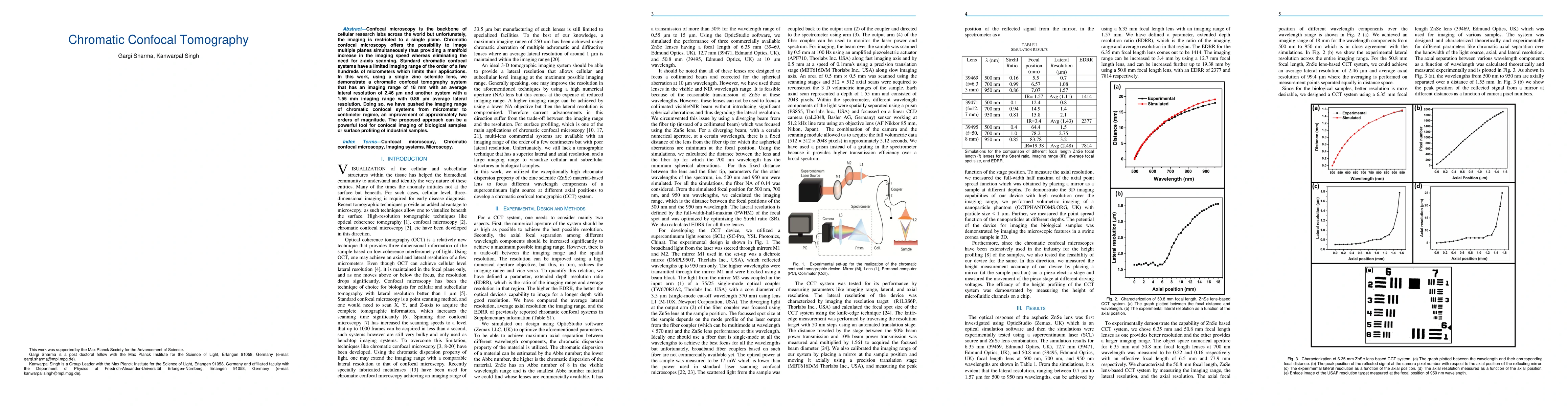
Confocal microscopy is the backbone of cellular research labs across the world but unfortunately, the imaging is restricted to a single plane. Chromatic confocal microscopy offers the possibility to image multiple planes simultaneously thus providing a manifold increase in the imaging speed whereas eliminating the need for z-axis scanning. Standard chromatic confocal systems have a limited imaging range of the order of a few hundreds of micrometers which limits their applications. In this work, using a single zinc selenide lens, we demonstrate a chromatic confocal tomography system that has an imaging range of 18 mm with an average lateral resolution of 2.46 microns and another system with a 1.55 mm imaging range with 0.86 microns average lateral resolution. Doing so, we have pushed the imaging range of chromatic confocal systems from micrometer to centimeter regime, an improvement of approximately two orders of magnitude. The proposed approach can be a powerful tool for confocal imaging of biological samples or surface profiling of industrial samples.
Seven facets of this paper, analysed and brought into focus by AI.
This research contributes to the development of advanced imaging techniques for biomedical applications, with potential implications for disease diagnosis and treatment.
A novel approach to chromatic confocal microscopy using a custom-designed lens system was employed.
This research contributes to the development of advanced imaging techniques for biomedical applications, with potential implications for disease diagnosis and treatment.
The development of a custom-designed lens system for chromatic confocal microscopy, which demonstrated improved image quality and depth of field compared to existing commercial systems.
This research presents a novel approach to chromatic confocal microscopy using a custom-designed lens system, offering improved performance and potential applications in biomedical imaging.
Current paper (gray), citations (green), references (blue)
Display is limited for performance on very large graphs.
Discussion 0