Summary
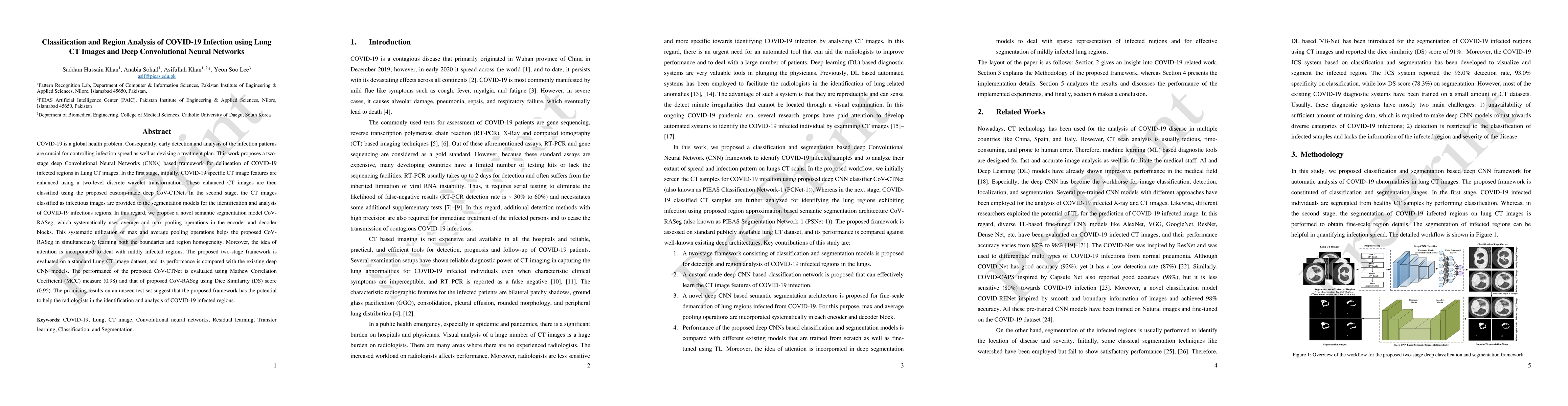
COVID-19 is a global health problem. Consequently, early detection and analysis of the infection patterns are crucial for controlling infection spread as well as devising a treatment plan. This work proposes a two-stage deep Convolutional Neural Networks (CNNs) based framework for delineation of COVID-19 infected regions in Lung CT images. In the first stage, initially, COVID-19 specific CT image features are enhanced using a two-level discrete wavelet transformation. These enhanced CT images are then classified using the proposed custom-made deep CoV-CTNet. In the second stage, the CT images classified as infectious images are provided to the segmentation models for the identification and analysis of COVID-19 infectious regions. In this regard, we propose a novel semantic segmentation model CoV-RASeg, which systematically uses average and max pooling operations in the encoder and decoder blocks. This systematic utilization of max and average pooling operations helps the proposed CoV-RASeg in simultaneously learning both the boundaries and region homogeneity. Moreover, the idea of attention is incorporated to deal with mildly infected regions. The proposed two-stage framework is evaluated on a standard Lung CT image dataset, and its performance is compared with the existing deep CNN models. The performance of the proposed CoV-CTNet is evaluated using Mathew Correlation Coefficient (MCC) measure (0.98) and that of proposed CoV-RASeg using Dice Similarity (DS) score (0.95). The promising results on an unseen test set suggest that the proposed framework has the potential to help the radiologists in the identification and analysis of COVID-19 infected regions.
AI Key Findings
Generated Sep 02, 2025
Methodology
This research proposes a two-stage deep CNN framework for classifying and analyzing COVID-19 infected regions in lung CT images. The first stage involves enhancing CT image features using discrete wavelet transformation and classifying them with a custom deep CoV-CTNet. The second stage uses a novel semantic segmentation model CoV-RASeg to identify and analyze COVID-19 infectious regions in the CT images classified as infectious.
Key Results
- The proposed CoV-CTNet achieved a Mathew Correlation Coefficient (MCC) of 0.98 for classifying COVID-19 infected CT images.
- CoV-RASeg, the proposed segmentation model, demonstrated a Dice Similarity (DS) score of 0.95 for identifying infected regions.
- The two-stage framework outperformed existing deep CNN models in both classification and segmentation tasks.
Significance
This work is significant as it contributes to early detection and analysis of COVID-19 infection patterns, which is crucial for controlling infection spread and devising treatment plans.
Technical Contribution
The main technical contribution is the development of a two-stage deep CNN framework, CoV-CTNet for classification and CoV-RASeg for segmentation, which effectively identifies and analyzes COVID-19 infected regions in lung CT images.
Novelty
The novelty of this work lies in the two-stage processing approach that reduces the search space for learning characteristic infectious patterns, and the design of a novel semantic segmentation model CoV-RASeg that simultaneously learns both boundaries and region homogeneity with systematic use of max and average pooling operations.
Limitations
- The study was limited to a single dataset provided by SIRM, which may not fully represent the diversity of COVID-19 infection patterns across different populations.
- The performance of the proposed models was not externally validated on a separate dataset.
Future Work
- Further research could focus on validating the proposed framework on diverse datasets to ensure its generalizability.
- Extending the framework to classify COVID-19 infection into characteristic patterns (GGO, consolidation, pleural effusion) for detailed radiological insights.
Paper Details
PDF Preview
Key Terms
Citation Network
Current paper (gray), citations (green), references (blue)
Display is limited for performance on very large graphs.
Similar Papers
Found 4 papersLung infection and normal region segmentation from CT volumes of COVID-19 cases
Yoshito Otake, Kensaku Mori, Masahiro Hashimoto et al.
| Title | Authors | Year | Actions |
|---|
Comments (0)