CNN-based fully automatic mitral valve extraction using CT images and existence probability maps
Publication
Metrics
AI Quick Summary
This paper proposes a fully automated method using DenseNet and U-Net to extract mitral valve shapes from CT images, improving accuracy over existing methods. The method uses both original CT images and existence probability maps, achieving a mean extraction error of 0.88 mm, outperforming traditional methods by 0.32 mm.
Paper Preview
Abstract
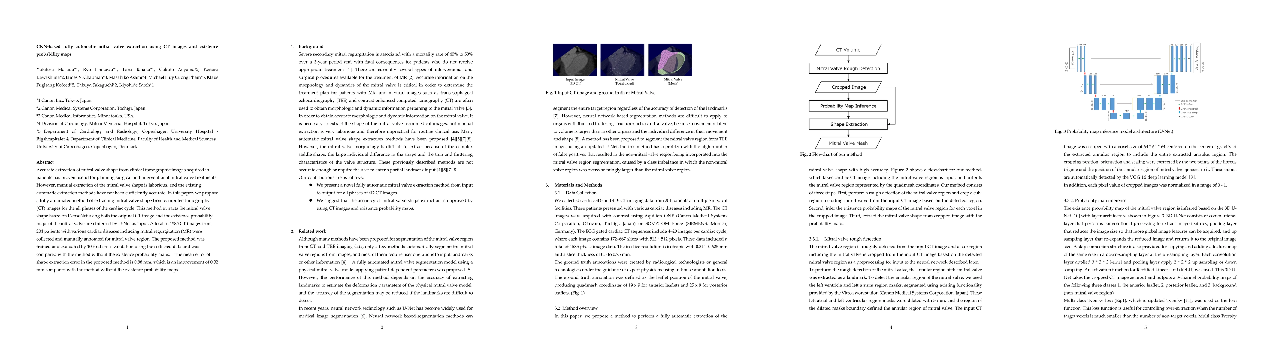
Accurate extraction of mitral valve shape from clinical tomographic images acquired in patients has proven useful for planning surgical and interventional mitral valve treatments. However, manual extraction of the mitral valve shape is laborious, and the existing automatic extraction methods have not been sufficiently accurate. In this paper, we propose a fully automated method of extracting mitral valve shape from computed tomography (CT) images for the all phases of the cardiac cycle. This method extracts the mitral valve shape based on DenseNet using both the original CT image and the existence probability maps of the mitral valve area inferred by U-Net as input. A total of 1585 CT images from 204 patients with various cardiac diseases including mitral regurgitation (MR) were collected and manually annotated for mitral valve region. The proposed method was trained and evaluated by 10-fold cross validation using the collected data and was compared with the method without the existence probability maps. The mean error of shape extraction error in the proposed method is 0.88 mm, which is an improvement of 0.32 mm compared with the method without the existence probability maps.
AI Key Findings
Get AI-generated insights about this paper's methodology, results, significance, and more — seven facets brought into focus.
Impact
Paper Details
Authors
PDF Preview
Key Terms
Citation Network
Current paper (gray), citations (green), references (blue)
Display is limited for performance on very large graphs.
Discussion 0