Publication
Metrics
AI Quick Summary
This study developed a novel imaging pipeline using scanning multiphoton microscopy to co-map cellular content and extracellular matrix with hemodynamics in intact arterial tissues, aiming to bridge the knowledge gap on how abnormal blood flow influences vascular pathologies. The methodology enables direct quantitative comparison of local flow and vascular wall biology in three dimensions.
Paper Preview
Abstract
Deviation of blood flow from an optimal range is known to be associated with the initiation and progression of vascular pathologies. Important open questions remain about how the abnormal flow drives specific wall changes in pathologies such as cerebral aneurysms where the flow is highly heterogeneous and complex. This knowledge gap precludes the clinical use of readily available flow data to predict outcomes and improve treatment of these diseases. As both flow and the pathological wall changes are spatially heterogeneous, a crucial requirement for progress in this area is a methodology for co-mapping local data from vascular wall biology with local hemodynamic data. In this study, we developed an imaging pipeline to address this pressing need. A protocol that employs scanning multiphoton microscopy was designed to obtain 3D data sets for smooth muscle actin, collagen and elastin in intact vascular specimens. A cluster analysis was developed to objectively categorize the smooth muscle cells (SMC) across the vascular specimen based on SMC density. In the final step in this pipeline, the location specific categorization of SMC, along with wall thickness was co-mapped with patient specific hemodynamic results, enabling direct quantitative comparison of local flow and wall biology in 3D intact specimens.
AI Key Findings
Get AI-generated insights about this paper's methodology, results, significance, and more — seven facets brought into focus.
Impact
Paper Details
Authors
PDF Preview
Key Terms
Citation Network
Current paper (gray), citations (green), references (blue)
Display is limited for performance on very large graphs.
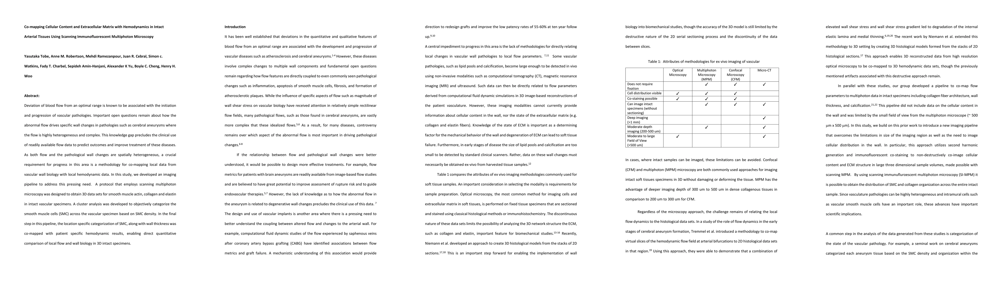
Discussion 0