Combined Diffusion-Relaxation MRI to Assess Muscle Microstructure and Composition
Publication
Metrics
Paper Preview
Abstract
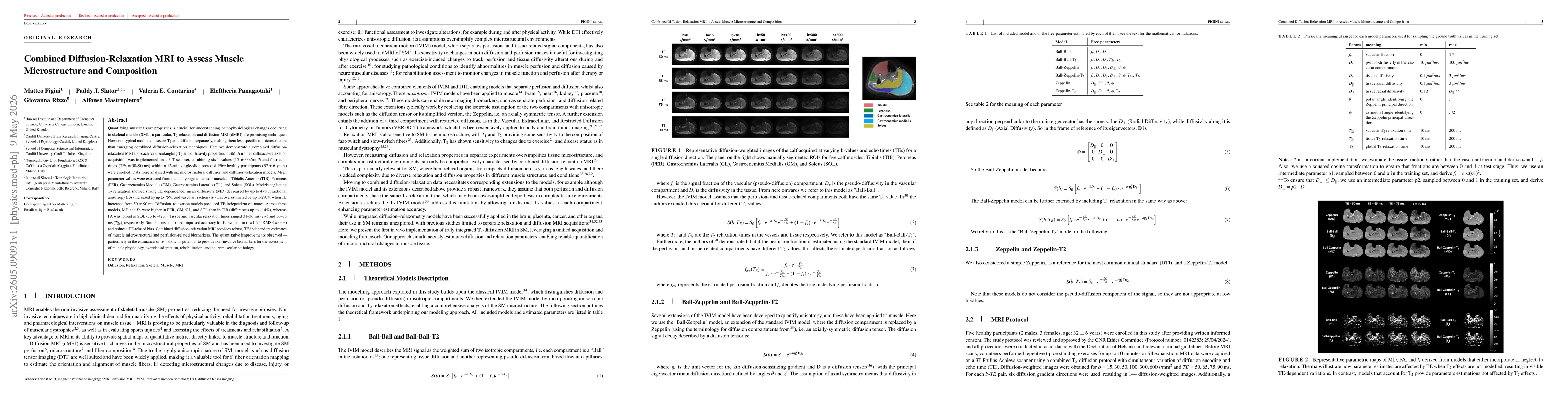
Quantifying muscle tissue properties is crucial for understanding pathophysiological changes occurring in skeletal muscle (SM). In particular, T2 relaxation and diffusion MRI (dMRI) are promising techniques. However, typical methods measure T2 and diffusion separately, making them less specific to microstructure than emerging combined diffusion-relaxation techniques. Here we demonstrate a combined diffusion-relaxation MRI approach for disentangling T2 and diffusivity properties in SM. A diffusion-relaxation acquisition was implemented on a 3 T scanner, combining six b-values and four echo times within a 12-min single-slice protocol. Five healthy participants were enrolled. Data were analysed with six microstructural diffusion and diffusion-relaxation models. Mean parameter values were extracted from manually segmented calf muscles. Models neglecting T2 relaxation showed strong TE dependence: mean diffusivity (MD) decreased by up to 47\%, fractional anisotropy (FA) increased by up to 75\%, and vascular fraction fv increased by up to 297\% when TE increased from 50 to 90 ms. Diffusion-relaxation models produced TE-independent estimates. Tissue and vascular relaxation times ranged 31-36 ms T2t and 66-86 ms T2v, respectively. Simulations confirmed improved accuracy for fv estimation (r=0.95; RMSE=0.03) and reduced TE-related bias. Combined diffusion-relaxation MRI provides robust, TE-independent estimates of muscle microstructural and perfusion-related biomarkers. The quantitative improvements observed - particularly in the estimation of fv - show its potential to provide non-invasive biomarkers for the assessment of muscle physiology, exercise adaptation, rehabilitation, and neuromuscular pathology.
AI Key Findings
Get AI-generated insights about this paper's methodology, results, significance, and more — seven facets brought into focus.
Discussion 0