Authors
Summary
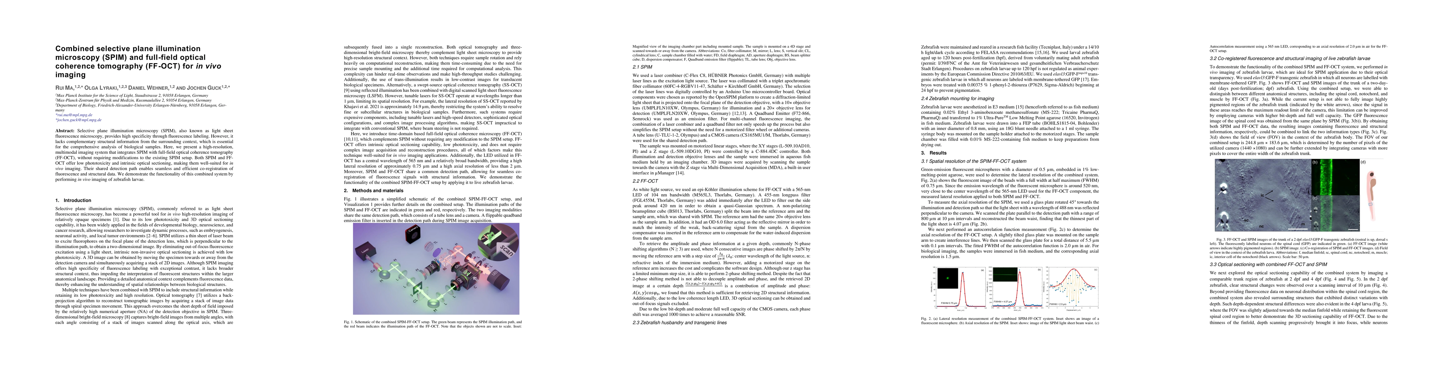
Selective plane illumination microscopy (SPIM), also known as light sheet fluorescence microscopy, provides high specificity through fluorescence labeling. However, it lacks complementary structural information from the surrounding context, which is essential for the comprehensive analysis of biological samples. Here, we present a high-resolution, multimodal imaging system that integrates SPIM with full-field optical coherence tomography (FF-OCT), without requiring modifications to the existing SPIM setup. Both SPIM and FF-OCT offer low phototoxicity and intrinsic optical sectioning, making them well-suited for in vivo imaging. Their shared detection path enables seamless and efficient co-registration of fluorescence and structural data. We demonstrate the functionality of this combined system by performing in vivo imaging of zebrafish larvae.
AI Key Findings
Get AI-generated insights about this paper's methodology, results, and significance.
Paper Details
PDF Preview
Similar Papers
Found 4 papersBond-Selective Full-Field Optical Coherence Tomography
Jian Zhao, Zian Wang, Fukai Chen et al.
Retinal blood flow imaging with combined full-field swept-source optical coherence tomography and laser Doppler holography
Léo Puyo, Clara Pfäffle, Hendrik Spahr et al.
No citations found for this paper.
Comments (0)