ArxivLens

Quick Summary:

This paper combines single-molecule super-resolved localization microscopy with fluorescence polarization imaging to simultaneously detect orthogonal polarization signals and localize proteins with super-resolution precision. The study demonstrates the potential of this dual-method approach to measure molecular stoichiometry and diffusion coefficients in biological samples.

Journal Reference: 2021 J. Phys. Photonics 3 034010
DOI: 10.1088/2515-7647/ac015d
arXiv Id: 2101.00048
Date Published: 2023-01-25
Date Updated: 2023-01-25

Key Terms (TF-IDF):

fluorescence (0.267)
polarization (0.226)
protein (0.198)
super (0.191)
biological (0.189)
cellular (0.189)
enable (0.187)
molecules (0.174)
resolved (0.174)
complexes (0.156)
processes (0.153)
simultaneous (0.151)
microscopy (0.150)
separate (0.143)
localization (0.141)
insights (0.127)
signals (0.125)
single (0.125)
imaging (0.124)
molecular (0.120)
License: http://arxiv.org/licenses/nonexclusive-distrib/1.0/

Summary:

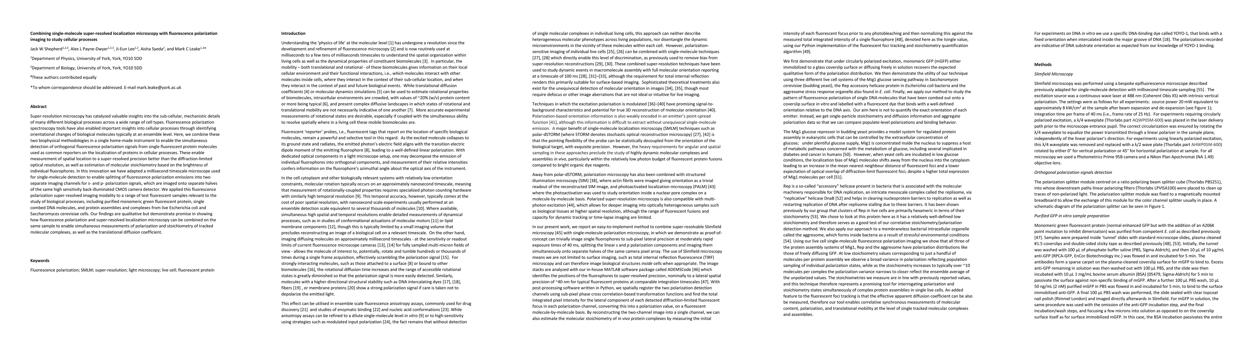
Super-resolution microscopy has catalyzed valuable insights into the sub-cellular, mechanistic details of many different biological processes across a wide range of cell types. Fluorescence polarization spectroscopy tools have also enabled important insights into cellular processes through identifying orientational changes of biological molecules typically at an ensemble level. Here, we combine these two biophysical methodologies in a single home-made instrument to enable the simultaneous detection of orthogonal fluorescence polarization signals from single fluorescent protein molecules used as common reporters on the localization of proteins in cellular processes. These enable measurement of spatial location to a super-resolved precision better than the diffraction-limited optical resolution, as well as estimation of molecular stoichiometry based on the brightness of individual fluorophores. In this innovation we have adapted a millisecond timescale microscope used for single-molecule detection to enable splitting of fluorescence polarization emissions into two separate imaging channels for s- and p- polarization signals, which are imaged onto separate halves of the same high sensitivity back-illuminated CMOS camera detector. We applied this fluorescence polarization super-resolved imaging modality to a range of test fluorescent samples relevant to the study of biological processes, including purified monomeric green fluorescent protein, single combed DNA molecules, and protein assemblies and complexes from live Escherichia coli and Saccharomyces cerevisiae cells. Our findings are qualitative but demonstrate promise in showing how fluorescence polarization and super-resolved localization microscopy can be combined on the same sample to enable simultaneous measurements of polarization and stoichiometry of tracked molecular complexes, as well as the translational diffusion coefficient.

Citation & Reference Network Visualization

This graph shows how this paper is connected to other research. The current paper is shown in dark gray, papers that cite it are in green, and papers it references are in blue. You can:

Current Paper
Papers citing this work (Citations)
Papers cited by this work (References)

• Hover over any node to see the paper title

• Click a node to view that paper's details

• Drag nodes to rearrange the graph

• Use the zoom controls (bottom right) to zoom in/out

Citations and References

Title Authors Year Actions
Illuminating cellular architecture and dynamics with fluorescence polarization microscopy.
W. F. Dean, A. Mattheyses
2024
View Paper
Cooperation between coagulase and von willebrand factor binding protein in Staphylococcus aureus fibrin pseudocapsule formation
Dominique C. S. Evans, A. Khamas, Alex L. Payne-Dwyer, A. Wollman, Kristian S. Rasmussen, J. Klitgaard, Birgitte Kallipolitis, M. Leake, R. Meyer
2024
View Paper
Bestrophin-like protein 4 is involved in photosynthetic acclimation to light fluctuations in Chlamydomonas
Liat Adler, C. S. Lau, K. Shaikh, Kim A van Maldegem, Alex L. Payne-Dwyer, Cécile Lefoulon, P. Girr, Nicky Atkinson, James Barrett, Tom Z Emrich-Mills, Emilija Dukic, Michael R. Blatt, M. Leake, Gilles Peltier, C. Spetea, Adrien Burlacot, Alistair J. McCormick, L. Mackinder, Charlotte E. Walker
2024
View Paper
SlimVar: rapid in vivo single-molecule tracking of chromatin regulators in plants
Alex L. Payne-Dwyer, Geng-Jen Jang, Caroline Dean, M. Leake
2024
View Paper
Predicting Rubisco-Linker Condensation from Titration in the Dilute Phase.
Alex L. Payne-Dwyer, Gaurav Kumar, James Barrett, Laura K. Gherman, Michael Hodgkinson, Michael J. Plevin, L. Mackinder, M. Leake, Charley Schaefer
2024
View Paper
Correlating fluorescence microscopy, optical and magnetic tweezers to study single chiral biopolymers such as DNA
J. Shepherd, Sebastien Guilbaud, Zhaokun Zhou, Jamieson A L Howard, Matthew Burman, Charley Schaefer, Adam M. Kerrigan, Clare Steele-King, A. Noy, M. Leake
2024
View Paper
A guide to small fluorescent probes for single-molecule biophysics
M. Leake, Steven D. Quinn
2023
View Paper
RecA and RecB: probing complexes of DNA repair proteins with mitomycin C in live Escherichia coli with single-molecule sensitivity
Alex L. Payne-Dwyer, A. Syeda, J. Shepherd, Lewis Frame, M. Leake
2021
View Paper
The End Restraint Method for Mechanically Perturbing Nucleic Acids In Silico.
J. Shepherd, M. Leake
2021
View Paper
Investigating molecular crowding during cell division in budding yeast with FRET
Sarah Lecinski, J. Shepherd, Lewis Frame, Imogen Hayton, C. MacDonald, M. Leake
2021
View Paper

Comments (0)

This website uses cookies to ensure you get the best experience. Learn more