Comparative Analysis of Unsupervised Algorithms for Breast MRI Lesion Segmentation
Publication
Metrics
AI Quick Summary
This paper compares three unsupervised algorithms—Gaussian Mixture Model, K-means clustering, and marker-controlled Watershed transformation—for segmenting breast lesions in MRI scans. The marker-controlled Watershed method showed superior performance in terms of segmentation accuracy, as evaluated by Dice similarity, Jaccard index, Hausdorff distance, and precision-recall metrics.
Paper Preview
Abstract
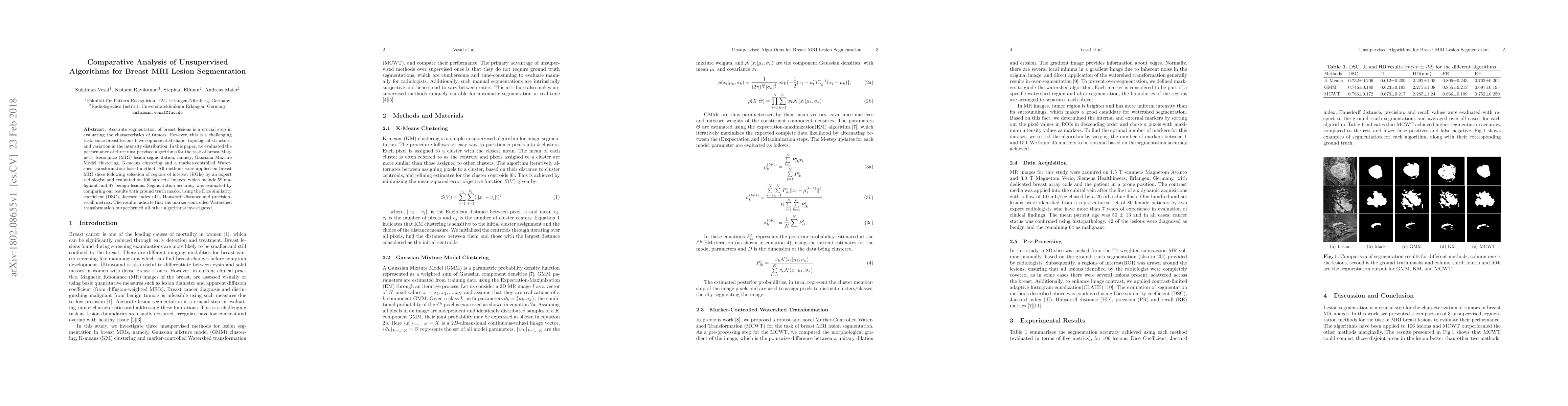
Accurate segmentation of breast lesions is a crucial step in evaluating the characteristics of tumors. However, this is a challenging task, since breast lesions have sophisticated shape, topological structure, and variation in the intensity distribution. In this paper, we evaluated the performance of three unsupervised algorithms for the task of breast Magnetic Resonance (MRI) lesion segmentation, namely, Gaussian Mixture Model clustering, K-means clustering and a marker-controlled Watershed transformation based method. All methods were applied on breast MRI slices following selection of regions of interest (ROIs) by an expert radiologist and evaluated on 106 subjects' images, which include 59 malignant and 47 benign lesions. Segmentation accuracy was evaluated by comparing our results with ground truth masks, using the Dice similarity coefficient (DSC), Jaccard index (JI), Hausdorff distance and precision-recall metrics. The results indicate that the marker-controlled Watershed transformation outperformed all other algorithms investigated.
AI Key Findings
Get AI-generated insights about this paper's methodology, results, significance, and more — seven facets brought into focus.
Impact
Paper Details
PDF Preview
Key Terms
Citation Network
Current paper (gray), citations (green), references (blue)
Display is limited for performance on very large graphs.
Discussion 0