Comparing Different Preprocessing Methods in Automated Segmentation of Retinal Vasculature
Publication
Metrics
AI Quick Summary
This study compares the effectiveness of two preprocessing methods—illumination equalization and contrast enhancement, and top-hat preprocessing—on retinal vessel segmentation using Laplacian-of-Gaussian, Canny, and matched filter edge detectors. Results show that preprocessing improves segmentation accuracy over 85% for all methods, with matched filter achieving the highest accuracy across different databases.
Paper Preview
Abstract
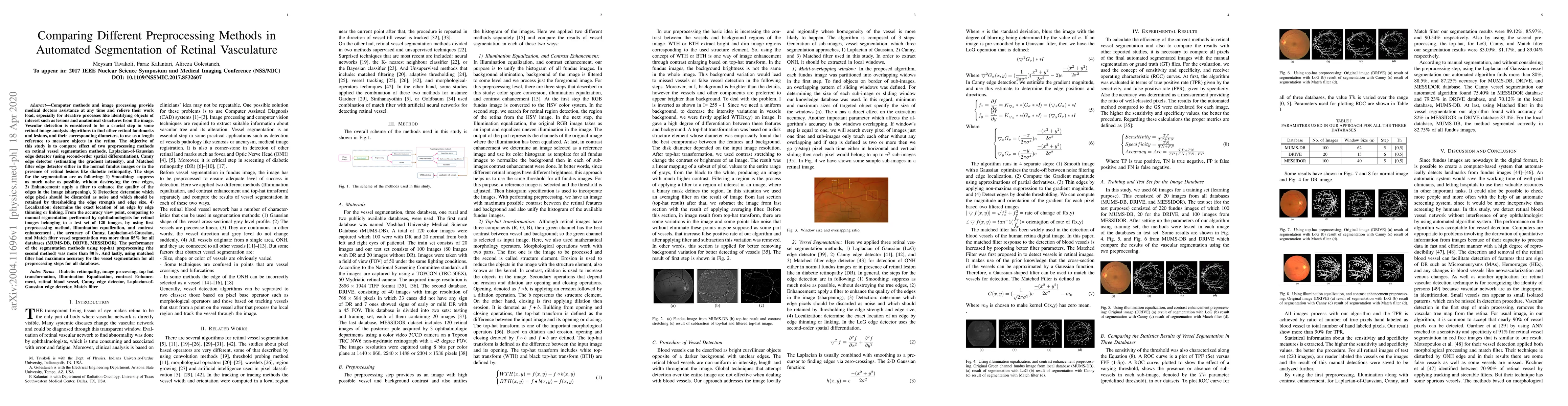
Computer methods and image processing provide medical doctors assistance at any time and relieve their workload, especially for iterative processes like identifying objects of interest such as lesions and anatomical structures from the image. Vascular detection is considered to be a crucial step in some retinal image analysis algorithms to find other retinal landmarks and lesions, and their corresponding diameters, to use as a length reference to measure objects in the retina. The objective of this study is to compare the effect of two preprocessing methods on retinal vessel segmentation methods, Laplacian-of-Gaussian edge detector (using second-order spatial differentiation), Canny edge detector (estimating the gradient intensity), and Matched filter edge detector either in the normal fundus images or in the presence of retinal lesions like diabetic retinopathy. From the accuracy viewpoint, compared to manual segmentation performed by ophthalmologists for retinal images belonging to a test set of 120 images, by using first preprocessing method, Illumination equalization, and contrast enhancement, the accuracy of Canny, Laplacian-of-Gaussian, and Match filter vessel segmentation was more than 85% for all databases (MUMS-DB, DRIVE, MESSIDOR). The performance of the segmentation methods using top-hat preprocessing (the second method) was more than 80%. And lastly, using matched filter had maximum accuracy for the vessel segmentation for all preprocessing steps for all databases.
AI Key Findings
Get AI-generated insights about this paper's methodology, results, significance, and more — seven facets brought into focus.
Impact
Paper Details
Authors
PDF Preview
Key Terms
Citation Network
Current paper (gray), citations (green), references (blue)
Display is limited for performance on very large graphs.
Discussion 0